
NucView® 530 Caspase-3 Substrate, 1 mM in 1X PBS
CAT:
37-10408
Size:
100 µL
Price:
Ask
- Availability: 24/48H Stock Items & 2 to 6 Weeks non Stock Items.
- Dry Ice Shipment: No


NucView® 530 Caspase-3 Substrate, 1 mM in 1X PBS
Description:
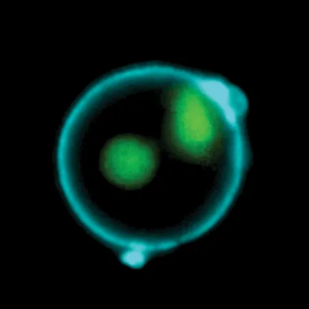
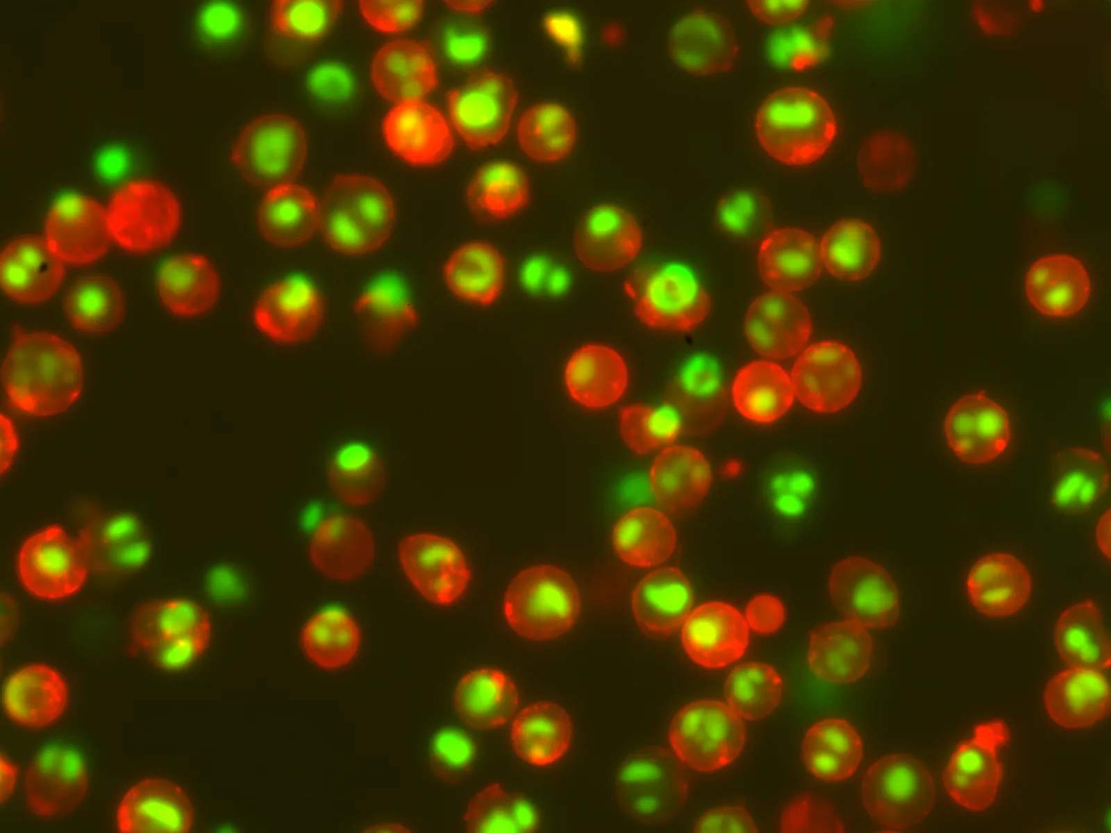
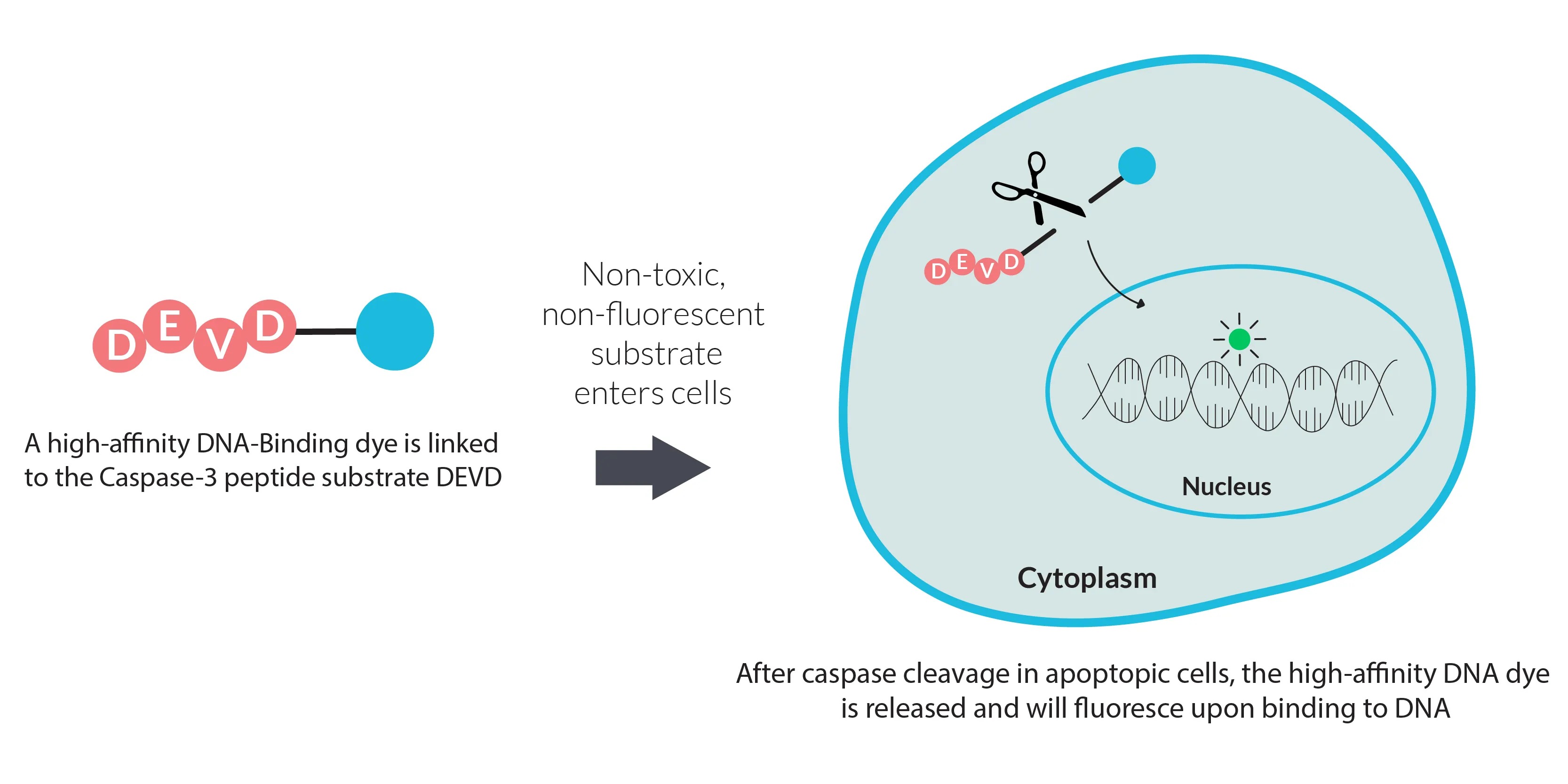
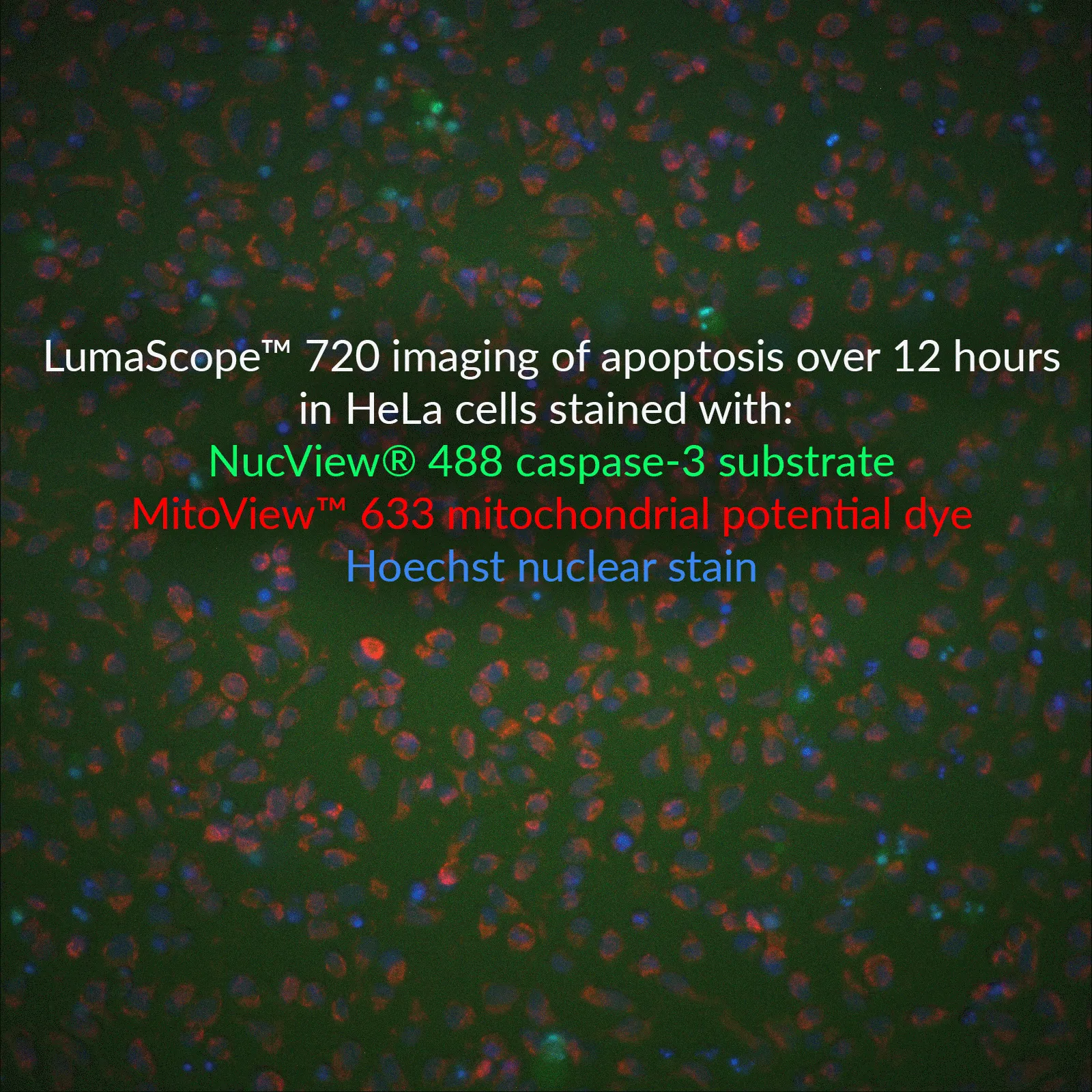
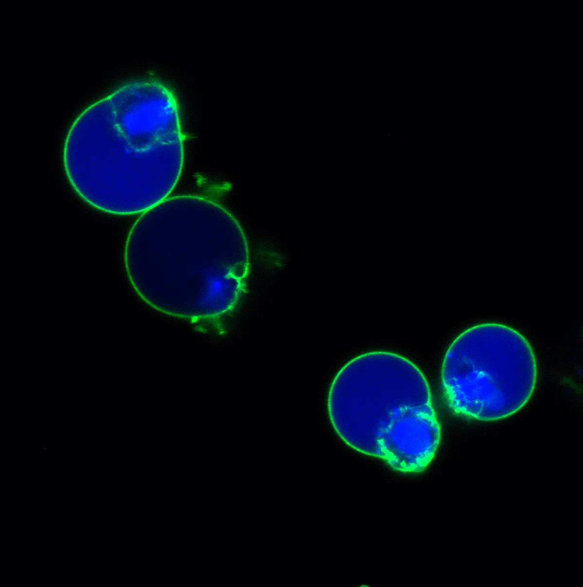
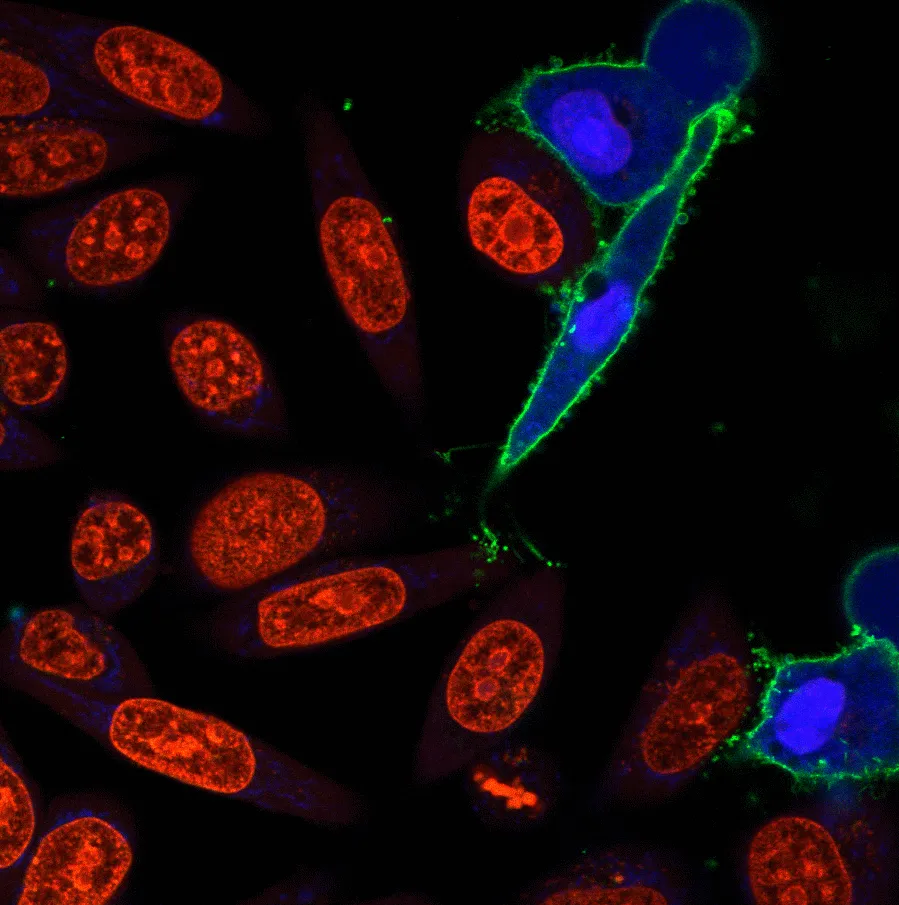
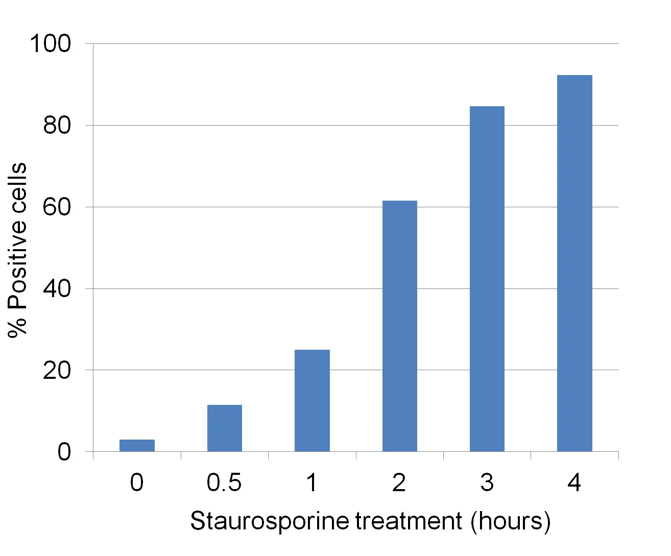
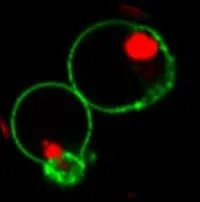
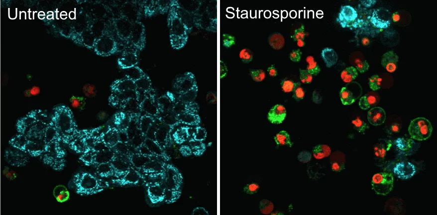
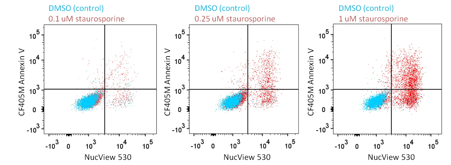
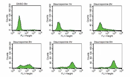
NucView® 530 Caspase-3 Substrate, 1 mM in PBS, provides a convenient tool for detecting apoptosis in intact cells based on caspase-3/7 activity using either confocal microscopy or flow cytometry. In contrast to other fluorogenic caspase substrates or fluorescent caspase inhibitor based (FLICA) assays, NucView® caspase-3 substrates can be used to detect caspase-3/7 activity within individual intact cells without inhibiting apoptosis progression. NucView® substrates consist of a fluorogenic DNA dye coupled to the caspase-3/7 DEVD recognition sequence. The substrate, which is initially non-fluorescent, penetrates the plasma membrane and enters the cytoplasm. In apoptotic cells, caspase-3/7 cleaves the substrate, releasing the high-affinity DNA dye, which migrates to the cell nucleus and stains DNA with fluorescence. Thus, NucView® Caspase-3 Substrates are bifunctional, allowing detection of caspase-3/7 activity and visualization of morphological changes in the nucleus during apoptosis. The staining is also formaldehyde-fixable. NucView® 530 Caspase-3 Substrate stains apoptotic cell nuclei with orange fluorescence (Ex/Em 528/563 nm), for detection in the Cy®3 channel by fluorescence microscopy (Figure 2) or the PE channel by flow cytometry. NucView® 530 can be used for multi-color imaging with blue, green, or far-red fluorescent probes. Note that when excited by the 488 nm laser line, NucView® 530 also fluoresces in the FITC channel, and therefore cannot be analyzed together with green probes by flow cytometry. NucView® 530 Caspase-3 Substrate also is available as a 1 mM solution in DMSO. The substrate in PBS is formulated for use in cells that are sensitive to DMSO toxicity. In non-DMSO sensitive cell types, adding DMSO during the substrate incubation may enhance NucView® 530 staining. Biotium also offers blue fluorogenic NucView® 405 Caspase-3 Substrate, and green fluorogenic NucView® 488 Caspase-3 Substrate and kits. Cy dye is a registered trademark of GE Healthcare.UNSPSC:
41116113UNSPSC Description:
Cytology reagents or solutions or stainsSource:
SyntheticStorage Conditions:
-35°C; Protect from lightShelf Life:
6 months
DATASHEET Document
View DocumentMSDS Document
View Document