
Anti-Catenin, beta (CTNNB1) (CTNNB1/2030R)
CAT:
37-BNUM2030-50
Size:
50 µL
Price:
Ask
- Availability: 24/48H Stock Items & 2 to 6 Weeks non Stock Items.
- Dry Ice Shipment: No
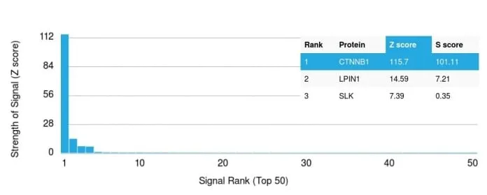


Anti-Catenin, beta (CTNNB1) (CTNNB1/2030R)
- Description: Beta-catenin associates with the cytoplasmic portion of E-cadherin. The catenin/cadherin complexes play an important role mediating cellular adhesion, including adherens junctions. Beta-catenin is involved in the Wnt signaling pathway as well as other signaling pathways, and is also found in complexes with many different proteins including the tumor suppressor protein APC. Defects in beta-catenin are associated with colorectal cancer, as well as many other cancer types. Beta-catenin is normally localized to the cell membrane, but can translocate to the nucleus in response to certain cell signaling. Primary antibodies are available purified, or with a selection of fluorescent CF® Dyes and other labels. CF® Dyes offer exceptional brightness and photostability. Note: Conjugates of blue fluorescent dyes like CF®405S and CF®405M are not recommended for detecting low abundance targets, because blue dyes have lower fluorescence and can give higher non-specific background than other dye colors.
- Synonyms: Beta-catenin; Catenin beta-1; Catenin (Cadherin associated protein), beta 1; CTNNB
- CAS Number: 9007-83-4
- UNSPSC: 41116161
- UNSPSC Description: Primary and secondary antibodies for multiple methodology immunostaining detection application
- Gene Name: CTNNB1
- Gene ID: 1499
- NCBI Gene ID: 476018
- UniProt: P35222
- Cellular Locus: Plasma membrane|Nucleus
- Host: Rabbit
- Species Reactivity: Human
- Immunogen: Recombinant full-length human β-catenin protein
- Target Antigen: Beta-catenin | Catenin, Beta
- Clonality: Recombinant Monoclonal
- Isotype: IgG
- Clone: CTNNB1/2030R
- Conjugation: Purified, BSA-free
- Disease: Tumor
- Source: Animal
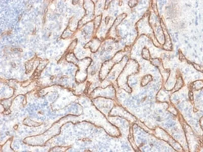
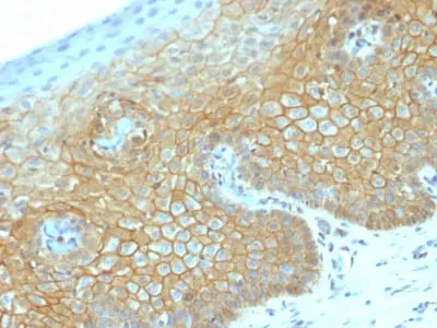
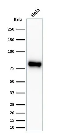
- Applications: Flow, intracellular (verified) | IF (verified) | IHC, FFPE (verified) | WB (verified)
- Validated Applications: FC, IF, IHC, FFPE, WB
- Field of Research: Cancer, Cell adhesion, Developmental biology, Signal transduction
- Positive Control: HeLa or MCF-7 cells. Breast carcinoma
- Concentration: 1 mg/mL
- Buffer: PBS, no BSA, no azide
- Molecular Weight: 92 kDa
- Additionnal Information: Higher concentration may be required for direct detection using primary antibody conjugates than for indirect detection with secondary antibody|Immunohistology (formalin): 1-2 ug/mL for 30 minutes at RT|Staining of formalin-fixed tissues requires boiling tissue sections in 10 mM citrate buffer, pH 6.0, for 10-20 minutes followed by cooling at RT for 20 minutes|Western blotting 1-2 ug/mL|Optimal dilution for a specific application should be determined by user
- Shipping Conditions: Room temperature
- Storage Conditions: -35°C to -5°C ; Stable at room temperature or 37°C (98°F) for 7 days.
- Shelf Life: 2 years