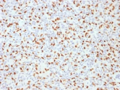
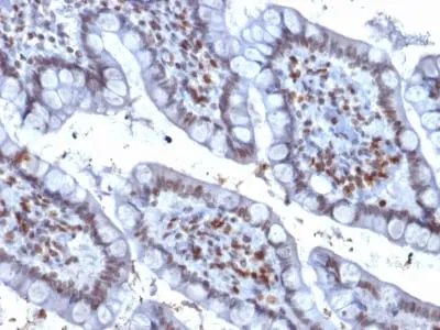
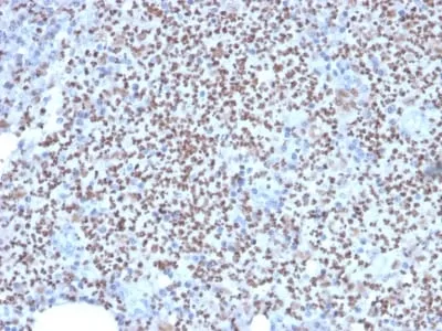
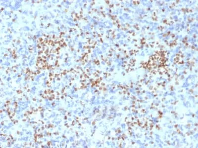
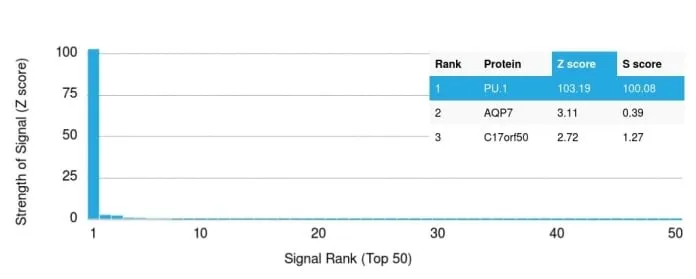
Anti-PU.1 (SPI-1) (B-Cell Marker) (PU1/2146), CF647 conjugate
CAT:
37-BNC472146-100
Size:
100 µL
Price:
Ask
- Availability: 24/48H Stock Items & 2 to 6 Weeks non Stock Items.
- Dry Ice Shipment: No


Anti-PU.1 (SPI-1) (B-Cell Marker) (PU1/2146), CF647 conjugate
Description:
PU.1 is a member of the ETS family of transcription factors and is important for normal B-cell development. It is expressed in the myeloid lineage and in immature as well as mature B-lymphocytes, with the exception of plasma cells. PU.1 is expressed in germinal center B-cells and mantle B-cells. Various lymphomas are also positive for this marker. It is essential during early B-cell differentiation. The absence of PU.1 results in total block of B-cell development at the pre-pro stage. PU.1 plays a key role in normal myeloid differentiation, and regulates the expression of immunoglobulin and other genes that are important for B-cell development._x000D_ _x000D_ Primary antibodies are available purified, or with a selection of fluorescent CF® Dyes and other labels. CF® Dyes offer exceptional brightness and photostability. Note: Conjugates of blue fluorescent dyes like CF®405S and CF®405M are not recommended for detecting low abundance targets, because blue dyes have lower fluorescence and can give higher non-specific background than other dye colors._x000D_ _x000D_Synonyms:
Transcription Factor spi1; 31kDa Transforming Protein; Hematopoietic Transcription Factor PU.1; SFPI1; SPI1; SPIA; Spleen focus forming virus (SFFV) proviral integration oncogene spi1UNSPSC:
41116161UNSPSC Description:
Primary and secondary antibodies for multiple methodology immunostaining detection applicationGene Name:
SPI1Gene ID:
6688NCBI Gene ID:
502511UniProt:
P17947Cellular Locus:
NucleusHost:
MouseSpecies Reactivity:
HumanImmunogen:
Recombinant fragment (around aa 16-170) of human PU.1 protein (Exact sequence is proprietary)Target Antigen:
PU.1Clonality:
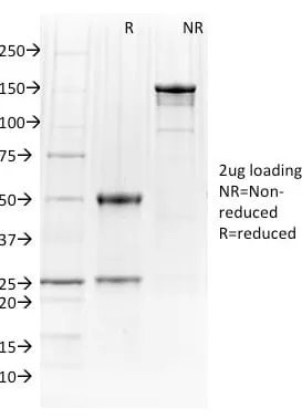
MonoclonalIsotype:
IgG2b κClone:
PU1/2146Conjugation:
CF647Source:
AnimalApplications:
Flow, intracellular (verified) | IF (verified) | IHC, FFPE (verified) | WB (verified)Validated Applications:
FC, IF, IHC, FFPE, WBField of Research:
Immunology, Transcription factorsPositive Control:
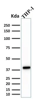
K-562 Cells, THP-1 cells. Lymph Node, Spleen, Hodgkin's Lymphoma, Colon Carcinoma.Concentration:
0.1 mg/mLBuffer:
PBS, 0.1% BSA, 0.05% azideMolecular Weight:
40 kDaAdditionnal Information:
Higher concentration may be required for direct detection using primary antibody conjugates than for indirect detection with secondary antibody|Immunohistology (formalin): 1-2 ug/mL for 30 minutes at RT|Western Blot 0.5-1 ug/mL|Staining of formalin-fixed tissues requires boiling tissue sections in 10 mM citrate buffer, pH 6.0, for 10-20 minutes followed by cooling at RT for 20 minutes|Optimal dilution for a specific application should be determined by userShipping Conditions:
Room temperatureStorage Conditions:
4°C; Protect from light; Stable at room temperature or 37°C (98°F) for 7 days.Shelf Life:
2 yearsCAS Number:
9007-83-4
DATASHEET Document
View DocumentMSDS Document
View Document