Anti-Ki-67 (Proliferating Cell Marker) (MKI67/2465), Biotin conjugate
CAT:
37-BNCB2465-100
Size:
100 µL
Price:
Ask
- Availability: 24/48H Stock Items & 2 to 6 Weeks non Stock Items.
- Dry Ice Shipment: No


Anti-Ki-67 (Proliferating Cell Marker) (MKI67/2465), Biotin conjugate
Description:
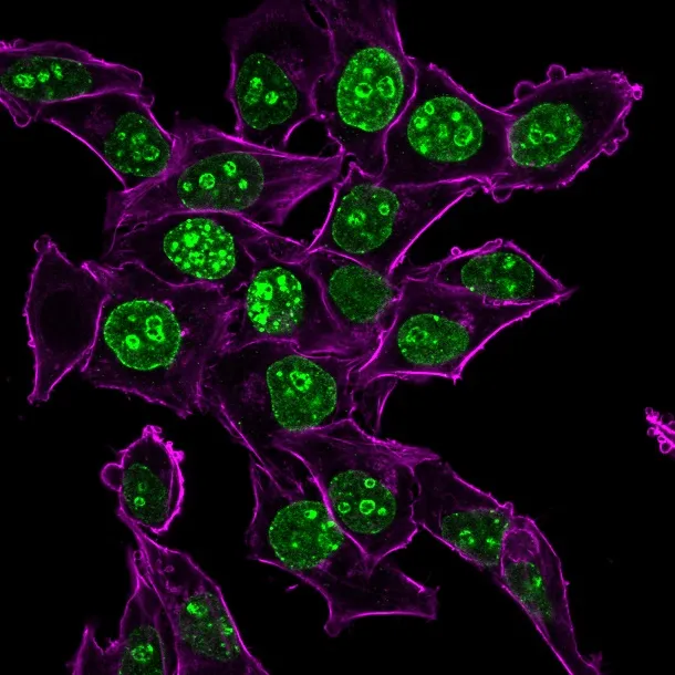
Ki-67 antigen is a nuclear, non-histone protein that is present in all stages of the cell cycle except G0. This characteristic makes Ki-67 an excellent marker for proliferating cells and is commonly used as one of the prognostic factors in cancer studies. A correlation has been demonstrated between Ki-67 index and the histo-pathological grade of neoplasms. Assessment of Ki-67 expression in renal and ureter tumors shows a correlation between tumor proliferation and disease progression, thus making it possible to differentiate high-risk patients. Ki-67 expression may also prove to be important for distinguishing between malignant and benign peripheral nerve sheath tumors. Ki-67 labeling index has been shown to be a prognostic marker in a number of neoplasms including grade II astrocytoma, oligodendroglioma, colon carcinoma, and breast carcinoma. In general, Ki-67 is a good marker of proliferating cell populations._x000D_ _x000D_ Primary antibodies are available purified, or with a selection of fluorescent CF® Dyes and other labels. CF® Dyes offer exceptional brightness and photostability. Note: Conjugates of blue fluorescent dyes like CF®405S and CF®405M are not recommended for detecting low abundance targets, because blue dyes have lower fluorescence and can give higher non-specific background than other dye colors._x000D_ _x000D_Synonyms:
KI-67; Ki67; KI-67 Antigen (KIA); MKI67; Proliferation related Ki-67 antigenUNSPSC:
41116161UNSPSC Description:
Primary and secondary antibodies for multiple methodology immunostaining detection applicationGene Name:
MKI67Gene ID:
4288NCBI Gene ID:
689823UniProt:
P46013Cellular Locus:
NucleusHost:
MouseSpecies Reactivity:
HumanImmunogen:
Recombinant fragment of human Ki67 protein (around aa 2293-2478) (exact sequence is proprietary)Target Antigen:
Ki-67Clonality:
MonoclonalIsotype:
IgG2b κClone:
MKI67/2465Conjugation:
BiotinSource:
AnimalApplications:
IF (verified) | IHC, FFPE (verified)Validated Applications:
IF, IHC, FFPEField of Research:
Cancer, Proliferating cell markersPositive Control:
Any actively proliferating cells. Skin, Tonsil or Lymph NodeConcentration:
0.1 mg/mLBuffer:
PBS, 0.1% BSA, 0.05% azideMolecular Weight:
345 kDa and 395 kDaAdditionnal Information:
Higher concentration may be required for direct detection using primary antibody conjugates than for indirect detection with secondary antibody|Immunohistology (formalin): 1-2 ug/mL for 30 minutes at RT|Staining of formalin-fixed tissues requires boiling tissue sections in 10 mM citrate buffer, pH 6.0, for 10-20 minutes followed by cooling at RT for 20 minutes|Optimal dilution for a specific application should be determined by userShipping Conditions:
Room temperatureStorage Conditions:
4°C; Stable at room temperature or 37°C (98°F) for 7 days.Shelf Life:
2 yearsCAS Number:
9007-83-4
DATASHEET Document
View DocumentMSDS Document
View Document