
Anti-TGFalpha(TGFA/1119), CF647 conjugate
- Availability: 24/48H Stock Items & 2 to 6 Weeks non Stock Items.
- Dry Ice Shipment: No


Anti-TGFalpha(TGFA/1119), CF647 conjugate
Description:
This antibody reacts with the C-terminus of TGF alpha and shows no cross-reaction with EGF and the neuropeptide synenkephalin. TGF-beta (aa50) is a growth factor with 33% homology to EGF, binds to EGFR, activates tyrosine phosphorylation of the receptor, and stimulates cell proliferation. It plays a role in tumor initiation by inducing the reversible transformed phenotype.Primary antibodies are available purified, or with a selection of fluorescent CF® Dyes and other labels. CF® Dyes offer exceptional brightness and photostability. Note: Conjugates of blue fluorescent dyes like CF®405S and CF®405M are not recommended for detecting low abundance targets, because blue dyes have lower fluorescence and can give higher non-specific background than other dye colors.Synonyms:
EGF-like TGF; ETGF; TFGA; TGF Type 1; TGFA; Wa1; Waved 1UNSPSC:
41116161UNSPSC Description:
Primary and secondary antibodies for multiple methodology immunostaining detection applicationGene Name:
TGFAGene ID:
7039NCBI Gene ID:
170009UniProt:
P01135Cellular Locus:
Secreted (extracellular)Host:
MouseSpecies Reactivity:
HumanImmunogen:
Recombinant human full-length TGF alpha proteinTarget Antigen:
TGF-AlphaClonality:
MonoclonalIsotype:
IgG1 κClone:
TGFA/1119Conjugation:
CF647Source:
AnimalApplications:
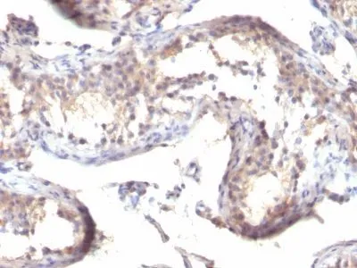
IHC, FFPE (verified)Validated Applications:
IHC, FFPEField of Research:
Signal transductionPositive Control:
Jurkat or Ramos cells. Testicular CarcinomaConcentration:
0.1 mg/mLBuffer:
PBS, 0.1% BSA, 0.05% azideMolecular Weight:
6 kDaAdditionnal Information:
Immunohistology formalin-paraffin 2-4 ug/mL|Staining of formalin-fixed tissues requires boiling tissue sections in 10 mM citrate buffer, pH 6.0, for 10-20 min followed by cooling at RT for 20 minutes|Flow Cytometry 0.5-1 ug/million cells/0.1 mL|Immunofluorescence 1-2 ug/mL|Optimal dilution for a specific application should be determined by userShipping Conditions:
Room temperatureStorage Conditions:
4°C; Protect from light; Stable at room temperature or 37°C (98°F) for 7 days.Shelf Life:
2 yearsCAS Number:
9007-83-4
DATASHEET Document
View DocumentMSDS Document
View Document