
Anti-Melanoma Marker(KBA.62)
CAT:
37-BNUB0895-100
Size:
100 µL
Price:
Ask
- Availability: 24/48H Stock Items & 2 to 6 Weeks non Stock Items.
- Dry Ice Shipment: No


Anti-Melanoma Marker(KBA.62)
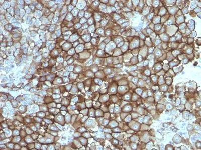
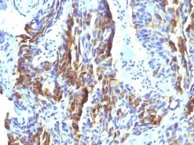
- Description: KBA.62 is a novel anti-melanoma antibody. It reacts positively against melanocytic tumors but not other tumors, thus demonstrating specificity and sensitivity. Moreover, it reacts positively against junctional nevus cells but not intradermal nevi, and against fetal melanocytes but not normal adult melanocytes. KBA.62 antibody is useful in identifying malignant melanomas. Metastatic amelanotic melanoma can often be confused with a variety of poorly differentiated carcinomas, large cell lymphomas, sarcomas, spindle cell carcinomas and various types of mesenchymal neoplasms. A keratin-negative, vimentin-rich neoplasm that immuno-reacts with antibody to S-100 protein and with KBA.62 antibody is, with rare exception, a melanoma. Anti-KBA.62 is a useful additional marker for melanoma, specifically in desmoplastic/spindle cell cases and in the context of micro-metastasis in sentinel lymph node.Primary antibodies are available purified, or with a selection of fluorescent CF® Dyes and other labels. CF® Dyes offer exceptional brightness and photostability. Note: Conjugates of blue fluorescent dyes like CF®405S and CF®405M are not recommended for detecting low abundance targets, because blue dyes have lower fluorescence and can give higher non-specific background than other dye colors.
- Synonyms: Human Melanoma Associated Antigen; KBA.62
- CAS Number: 9007-83-4
- UNSPSC: 41116161
- UNSPSC Description: Primary and secondary antibodies for multiple methodology immunostaining detection application
- Gene ID: Not Known
- NCBI Gene ID: Not Known
- UniProt: Not Known
- Cellular Locus: Cytoplasmic|Plasma membrane
- Host: Mouse
- Species Reactivity: Human
- Immunogen: Human KAL cells derived from lymph node metastasis of malignant melanoma
- Target Antigen: Melanoma Marker
- Clonality: Monoclonal
- Isotype: IgG1 κ
- Clone: KBA.62
- Conjugation: Purified, with BSA
- Disease: Tumor
- Source: Animal
- Applications: IHC, FFPE (verified)
- Validated Applications: IHC, FFPE
- Field of Research: Cancer
- Positive Control: SK-MEL-13 and SK-MEL-19 Melanoma cell lines; Melanomas
- Concentration: 0.2 mg/mL
- Buffer: PBS, 0.05% BSA, 0.05% azide
- Molecular Weight: Multiple (140, 135 and 128 kDa and two weak bands of 88 and 73 kDa)
- Additionnal Information: Higher concentration may be required for direct detection using primary antibody conjugates than for indirect detection with secondary antibody|Immunofluorescence: 0.5-1 ug/mL|Immunohistology (formalin)|Staining of formalin-fixed tissues requires boiling tissue sections in 10 mM citrate buffer, pH 6.0, for 10-20 min followed by cooling at RT for 20 minutes|Optimal dilution for a specific application should be determined by user
- Shipping Conditions: Room temperature
- Storage Conditions: 4°C; Stable at room temperature or 37°C (98°F) for 7 days.
- Shelf Life: 2 years