
Anti-Cdc20(CDC20/1102), CF740 conjugate
CAT:
37-BNC741102-100
Size:
100 µL
Price:
Ask
- Availability: 24/48H Stock Items & 2 to 6 Weeks non Stock Items.
- Dry Ice Shipment: No


Anti-Cdc20(CDC20/1102), CF740 conjugate
Description:
Cyclins, regulatory subunits that associate with kinases, control many of the important steps in cell cycle progression. The Cdc2 protein kinase (p34Cdc2) exhibits protein kinase activity in vitro and exists in a complex with both cyclin B and a protein homologous to p13SUC1. Cdc2 kinase is the active subunit of the M phase promoting factor (MPF) and the M phase-specific Histone H1 kinase. The p34Cdc2/cyclin B complex is required for the G2 to M transition. An additional cell cycle-dependent protein kinase, termed p55cdc, exhibits a high degree of homology with the S. cerevisiae proteins Cdc20 and Cdc4. The p55cdc transcript is readily detectable in a variety of cultured cell lines in growth phase, but disappears when cell growth is chemically arrested.Primary antibodies are available purified, or with a selection of fluorescent CF® Dyes and other labels. CF® Dyes offer exceptional brightness and photostability. Note: Conjugates of blue fluorescent dyes like CF®405S and CF®405M are not recommended for detecting low abundance targets, because blue dyes have lower fluorescence and can give higher non-specific background than other dye colors.Synonyms:
CDC20, CDC20A, p55CDC, P55CDC-LSBUNSPSC:
41116161UNSPSC Description:
Primary and secondary antibodies for multiple methodology immunostaining detection applicationGene Name:
CDC20Gene ID:
991NCBI Gene ID:
524947UniProt:
Q12834Cellular Locus:
Cytoplasmic|NucleusHost:
MouseSpecies Reactivity:
HumanImmunogen:
Recombinant human Cdc20 proteinTarget Antigen:
Cdc20Clonality:
MonoclonalIsotype:
IgG1 κClone:
CDC20/1102Conjugation:
CF740Source:
AnimalApplications:
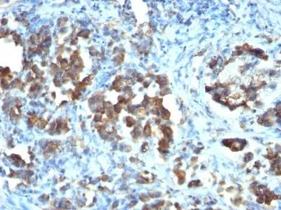
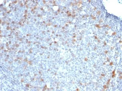
IHC, FFPE (verified)Validated Applications:
IHC, FFPEField of Research:
Cell cyclePositive Control:
Ramos or HeLa cells. Tonsil or gastric carcinoma.Concentration:
0.1 mg/mLBuffer:
PBS, 0.1% rBSA, 0.05% azideMolecular Weight:
55 kDaAdditionnal Information:
Immunohistology formalin-fixed 2-4 ug/mL|Staining of formalin-fixed tissues requires boiling tissue sections in 10 mM citrate buffer, pH 6.0, for 10-20 min followed by cooling at RT for 20 minutes|Immunofluorescence 0.5-1 ug/mL|Flow Cytometry 0.5-1 ug/million cells/0.1 mL|Optimal dilution for a specific application should be determined by userShipping Conditions:
Room temperatureStorage Conditions:
4°C; Protect from light; Stable at room temperature or 37°C (98°F) for 7 days.Shelf Life:
2 yearsCAS Number:
9007-83-4
DATASHEET Document
View DocumentMSDS Document
View Document