Anti-Cytokeratin, multi(C11)
CAT:
37-BNUB0393-500
Size:
500 µL
Price:
Ask
- Availability: 24/48H Stock Items & 2 to 6 Weeks non Stock Items.
- Dry Ice Shipment: No
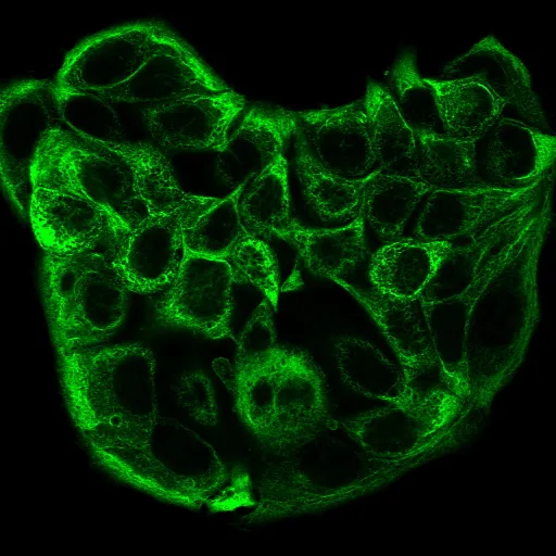
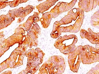


Anti-Cytokeratin, multi(C11)
Description:
This MAb recognizes cytokeratin 4, 5, 6, 8, 10, 13, and 18. This is a broad-spectrum antibody, which has been reported to differentiate epithelial tumors from non-epithelial tumors. Many studies have shown the usefulness of keratins as markers in cancer research and tumor diagnosis._x000D_ _x000D_ Primary antibodies are available purified, or with a selection of fluorescent CF® Dyes and other labels. CF® Dyes offer exceptional brightness and photostability. Note: Conjugates of blue fluorescent dyes like CF®405S and CF®405M are not recommended for detecting low abundance targets, because blue dyes have lower fluorescence and can give higher non-specific background than other dye colors._x000D_ _x000D_Synonyms:
K1B; KRT1B; Keratin, type II cytoskeletal 1b; K77; CK-1B; Keratin 1B; Keratin-77; Cytokeratin-1B; Type-II Keratin Kb39UNSPSC:
41116161UNSPSC Description:
Primary and secondary antibodies for multiple methodology immunostaining detection applicationGene Name:
KRT4Gene ID:
3851 (CK4); 3852 (CK5); 3853 (CK6A); 3854 (CK6B); 286887 (CK6C); 3856 (CK8); 3858 (CK10); 3860 (CK13); 3875 (CK18)UniProt:
P19013 (CK4); P13647 (CK5); P02538 (CK6A); P04259 (CK6B); P48668 (CK6C); P05787 (CK8); P13645 (CK10); P13646 (CK13); P05783 (CK18)Cellular Locus:
CytoskeletonHost:
MouseSpecies Reactivity:
Cow, Frog, Goat, Guinea pig, Human, Marmoset, Mouse, Pig, RatImmunogen:
Keratin-enriched preparation from cultured human A431Target Antigen:
Cytokeratin, multiClonality:
MonoclonalIsotype:
IgG1Clone:
C11Conjugation:
Purified, with BSASource:
AnimalApplications:
Flow, intracellular (verified) | IF (verified) | IHC, FFPE (verified) | WB (published)Validated Applications:
FC, IF, IHC, FFPE, WBField of Research:
Cancer, CytoskeletonPositive Control:
A431 cells, Skin, Colon carcinomaConcentration:
0.2 mg/mLBuffer:
PBS, 0.05% BSA, 0.05% azideMolecular Weight:
MultipleAdditionnal Information:
Higher concentration may be required for direct detection using primary antibody conjugates than for indirect detection with secondary antibody|Immunohistology formalin-fixed 0.5-1.0ug/mL|Staining of formalin-fixed tissues requires boiling tissue sections in 10 mM citrate buffer, pH 6.0, for 10-20 minutes followed by cooling at RT for 20 minutes|Flow Cytometry 0.5-1 ug/million cells/0.1 mL|Immunofluorescence 0.5-1.0 ug/mL|Western blotting 0.5-1.0 ug/mL|Optimal dilution for a specific application should be determined by userReferences & Citations:
Note: References for this clone sold by other suppliers may be listed for expected applications. Clin Cancer Res (2012) 18(4): 993-1003. (IHC, FFPE; western)Shipping Conditions:
Room temperatureStorage Conditions:
4°C; Stable at room temperature or 37°C (98°F) for 7 days.Shelf Life:
2 yearsCAS Number:
9007-83-4
DATASHEET Document
View DocumentMSDS Document
View Document