Anti-TACSTD2 / TROP2 (Epithelial Marker) (TACSTD2/2151), CF740 conjugate
CAT:
37-BNC742151-100
Size:
100 µL
Price:
Ask
- Availability: 24/48H Stock Items & 2 to 6 Weeks non Stock Items.
- Dry Ice Shipment: No


Anti-TACSTD2 / TROP2 (Epithelial Marker) (TACSTD2/2151), CF740 conjugate
Description:
TACSTD2 is a cell surface glycoprotein receptor. It is a single pass type I membrane protein containing one thyroglobulin type-1 domain, an epidermal growth factor-like repeat, a phosphatidylinositol binding site and tyrosine phosphorylation sites near the C-terminus. It plays a role in transducing intracellular calcium signals. It is expressed in trophoblast cells, cornea and multi-stratified epithelia. It is also highly expressed in several types of tumors and is involved in regulating the growth of carcinoma cells.Primary antibodies are available purified, or with a selection of fluorescent CF® Dyes and other labels. CF® Dyes offer exceptional brightness and photostability. Note: Conjugates of blue fluorescent dyes like CF®405S and CF®405M are not recommended for detecting low abundance targets, because blue dyes have lower fluorescence and can give higher non-specific background than other dye colors.Synonyms:
Cell surface glycoprotein Trop-2; Membrane Component Chromosome 1, Surface Marker 1 (M1S1); Pancreatic Carcinoma Marker Protein GA733-1; TROP2; Tumor-Associated Calcium Signal Transducer 2 (TACSTD2)CAS Number:
9007-83-4UNSPSC:
41116161UNSPSC Description:
Primary and secondary antibodies for multiple methodology immunostaining detection applicationGene Name:
TACSTD2Gene ID:
4070NCBI Gene ID:
23582UniProt:
P09758Cellular Locus:
Plasma membraneHost:
MouseSpecies Reactivity:
HumanImmunogen:
Recombinant fragment of human TACSTD2 protein (around aa 31-274) (exact sequence is proprietary)Target Antigen:
TACSTD2 | TROP2Clonality:
MonoclonalIsotype:
IgG2b κClone:
TACSTD2/2151Conjugation:
CF740Source:
AnimalApplications:
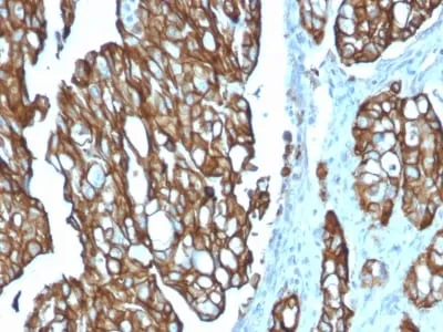
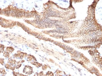
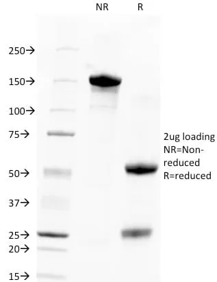
IHC, FFPE (verified) | WB (verified)Validated Applications:
IHC, FFPE, WBField of Research:
Cancer, Signal transductionPositive Control:
HT29 cells. Breast or Colon CarcinomaConcentration:
0.1 mg/mLBuffer:
PBS, 0.1% rBSA, 0.05% azideMolecular Weight:
40 kDaAdditionnal Information:
Higher concentration may be required for direct detection using primary antibody conjugates than for indirect detection with secondary antibody|Immunohistology (formalin): 1-2 ug/mL for 30 minutes at RT|Staining of formalin-fixed tissues requires boiling tissue sections in 10 mM citrate buffer, pH 6.0, for 10-20 minutes followed by cooling at RT for 20 minutes|Western blotting 0.5-1 ug/mL|Optimal dilution for a specific application should be determined by userShipping Conditions:
Room temperatureStorage Conditions:
4°C; Protect from light; Stable at room temperature or 37°C (98°F) for 7 days.Shelf Life:
2 years
DATASHEET Document
View DocumentMSDS Document
View Document