Anti-Arginase 1 (Hepatocellular Carcinoma Marker) (ARG1/1125) , CF640R conjugate
CAT:
37-BNC401125-100
Size:
100 µL
Price:
Ask
- Availability: 24/48H Stock Items & 2 to 6 Weeks non Stock Items.
- Dry Ice Shipment: No


Anti-Arginase 1 (Hepatocellular Carcinoma Marker) (ARG1/1125) , CF640R conjugate
Description:
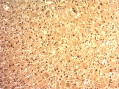
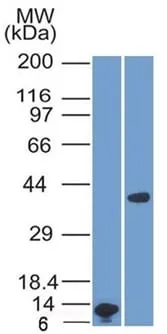
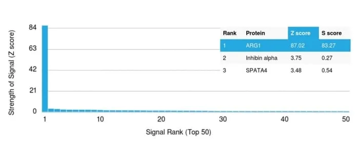
This antibody recognizes a protein of 35-38 kDa, which is identified as Arginase 1 (ARG1). Arginase is a manganese metallo-enzyme that catalyzes the hydrolysis of arginine to generate ornithine and urea. Arginase I and II are isoenzymes which differ in subcellular localization, regulation, and possibly function. Arginase I is a cytosolic enzyme, which is expressed mainly in the liver as part of the urea cycle, whereas arginase II is a mitochondrial protein found in a variety of tissues. Antibodies to Arginase 1 label hepatocytes in normal tissues and granulocytes in peripheral blood. Arginase 1 is a sensitive and specific marker for identification of hepatocellular carcinoma.Primary antibodies are available purified, or with a selection of fluorescent CF® Dyes and other labels. CF® Dyes offer exceptional brightness and photostability. Note: Conjugates of blue fluorescent dyes like CF®405S and CF®405M are not recommended for detecting low abundance targets, because blue dyes have lower fluorescence and can give higher non-specific background than other dye colors.Synonyms:
Arginase 1; ARG1; liver-type arginase; type I arginaseUNSPSC:
41116161UNSPSC Description:
Primary and secondary antibodies for multiple methodology immunostaining detection applicationGene Name:
ARG1Gene ID:
383NCBI Gene ID:
440934UniProt:
P05089Cellular Locus:
CytoplasmicHost:
MouseSpecies Reactivity:
HumanImmunogen:
Recombinant human ARG1 protein fragment (around aa11-97) (exact sequence is proprietary)Target Antigen:
Arginase 1Clonality:
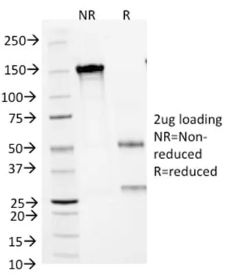
MonoclonalIsotype:
IgG3 κClone:
ARG1/1125Conjugation:
CF640RSource:
AnimalApplications:
IHC, FFPE (verified) | WB (verified)Validated Applications:
IHC, FFPE, WBField of Research:
Cancer, MetabolismPositive Control:
293Tcells. Hepatocellular Carcinoma (HCC).Concentration:
0.1 mg/mLBuffer:
PBS, 0.1% BSA, 0.05% azideMolecular Weight:
35-38 kDaAdditionnal Information:
Higher concentration may be required for direct detection using primary antibody conjugates than for indirect detection with secondary antibody|Immunohistology (formalin): 2-4 ug/mL for 30 minutes at RT|Staining of formalin-fixed tissues requires boiling tissue sections in 10 mM citrate buffer, pH 6.0, for 10-20 minutes followed by cooling at RT for 20 minutes|Western blotting 1-2 ug/mL|Optimal dilution for a specific application should be determined by userShipping Conditions:
Room temperatureStorage Conditions:
4°C; Protect from light; Stable at room temperature or 37°C (98°F) for 7 days.Shelf Life:
2 yearsCAS Number:
9007-83-4
DATASHEET Document
View DocumentMSDS Document
View Document