
Anti-Tyrosinase (Melanoma Marker) (TYR/2024R)
- Availability: 24/48H Stock Items & 2 to 6 Weeks non Stock Items.
- Dry Ice Shipment: No


Anti-Tyrosinase (Melanoma Marker) (TYR/2024R)
Description:
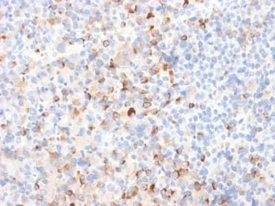
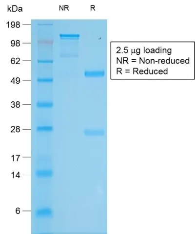
This antibody recognizes a cluster of proteins between 70-80 kDa, identified as tyrosinase. Occasionally a minor band at 55 kDa is also detected. This MAb shows no cross-reaction with MAGE-1 and tyrosinase-related protein 1, TRP-1/gp75. Tyrosinase is a copper-containing metalloglycoprotein that catalyzes several steps in the melanin pigment biosynthetic pathway; the hydroxylation of tyrosine to L-3,4-dihydroxy-phenylalanine (dopa), and the subsequent oxidation of dopa to dopaquinone. Mutations of the tyrosinase gene occur in various forms of albinism. Tyrosinase is one of the targets for cytotoxic T-cell recognition in melanoma patients. Staining of melanomas with this MAb shows tyrosinase in melanotic as well as amelanotic variants. This MAb is a useful marker for melanocytes and melanomas.Primary antibodies are available purified, or with a selection of fluorescent CF® Dyes and other labels. CF® Dyes offer exceptional brightness and photostability. Note: Conjugates of blue fluorescent dyes like CF®405S and CF®405M are not recommended for detecting low abundance targets, because blue dyes have lower fluorescence and can give higher non-specific background than other dye colors.Synonyms:
ATN, CMM8, LB24-AB, Monophenol monooxygenase, OCA1, OCA1A, Oculocutaneous albinism IA, SHEP3, SK29-AB, Tumor rejection antigen AB, TYRUNSPSC:
41116161UNSPSC Description:
Primary and secondary antibodies for multiple methodology immunostaining detection applicationGene Name:
TYRGene ID:
7299NCBI Gene ID:
503555UniProt:
P14679Cellular Locus:
VesicularHost:
RabbitSpecies Reactivity:
HumanImmunogen:
Recombinant full-length human tyrosinase (TYR) proteinTarget Antigen:
TyrosinaseClonality:
Recombinant MonoclonalIsotype:
IgGClone:
TYR/2024RConjugation:
Purified, BSA-freeDisease:
TumorSource:
AnimalApplications:
IHC, FFPE (verified)Validated Applications:
IHC, FFPEField of Research:
CancerPositive Control:
SK-MEL-19, SK-MEL-30 cells or Melanoma.Concentration:
1 mg/mLBuffer:
PBS, no BSA, no azideMolecular Weight:
70-80 kDaAdditionnal Information:
Higher concentration may be required for direct detection using primary antibody conjugates than for indirect detection with secondary antibody|Immunohistology (formalin): 0.5-1 ug/mL for 30 minutes at RT|Staining of formalin-fixed tissues requires boiling tissue sections in 10 mM Tris with 1 mM EDTA pH 9.0 or 10 mM citrate buffer pH6.0 for 10-20 minutes followed by cooling at RT for 20 minutes|Optimal dilution for a specific application should be determined by userShipping Conditions:
Room temperatureStorage Conditions:
-35°C to -5°C ; Stable at room temperature or 37°C (98°F) for 7 days.Shelf Life:
2 yearsCAS Number:
9007-83-4
DATASHEET Document
View DocumentMSDS Document
View Document