Anti-Emerin (Papillary Thyroid Carcinoma and EDMD Marker) (EMD/2167), CF640R conjugate
CAT:
37-BNC402167-100
Size:
100 µL
Price:
Ask
- Availability: 24/48H Stock Items & 2 to 6 Weeks non Stock Items.
- Dry Ice Shipment: No


Anti-Emerin (Papillary Thyroid Carcinoma and EDMD Marker) (EMD/2167), CF640R conjugate
Description:
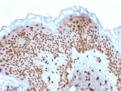
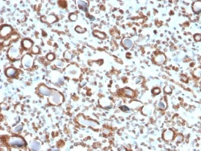
Emerin is a member of the nuclear lamina associated protein family. It is ubiquitously expressed and localized to the nuclear membrane in normal cells. Mutations of the gene that encodes emerin result in the X-linked recessive disease Emery-Dreifuss muscular dystrophy (EDMD), which is characterized by slowly progressing contractures, skeletal muscle wasting and cardiomyopathy. Reportedly, lack of Emerin expression is one cause of EDMD. Emerin is involved in the association of the nuclear membrane with the lamina, and is localized specifically to desmosomes and fasciae adherents in the heart. Identification of nuclear membrane irregularities with anti-emerin antibody has been reported useful in diagnosing papillary thyroid carcinoma.Primary antibodies are available purified, or with a selection of fluorescent CF® Dyes and other labels. CF® Dyes offer exceptional brightness and photostability. Note: Conjugates of blue fluorescent dyes like CF®405S and CF®405M are not recommended for detecting low abundance targets, because blue dyes have lower fluorescence and can give higher non-specific background than other dye colors.Synonyms:
EMD; Emerin; Emery Dreifuss muscular dystrophy (EDMD); STAUNSPSC:
41116161UNSPSC Description:
Primary and secondary antibodies for multiple methodology immunostaining detection applicationGene Name:
EMDGene ID:
2010NCBI Gene ID:
522823UniProt:
P50402Cellular Locus:
Nuclear membraneHost:
MouseSpecies Reactivity:
HumanImmunogen:
Recombinant human Emerin protein fragment (around aa 56-167) (exact sequence is proprietary)Target Antigen:
EmerinClonality:
MonoclonalIsotype:
IgG1Clone:
EMD/2167Conjugation:
CF640RSource:
AnimalApplications:
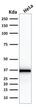
IF (verified) | IHC, FFPE (verified) | WB (verified)Validated Applications:
IF, IHC, FFPE, WBField of Research:
Organelle markersPositive Control:
K-562, HeLa or Jurkat cells. Kidney or SkinConcentration:
0.1 mg/mLBuffer:
PBS, 0.1% BSA, 0.05% azideMolecular Weight:
37 kDaAdditionnal Information:
Higher concentration may be required for direct detection using primary antibody conjugates than for indirect detection with secondary antibody|Immunohistology (formalin): 1-2 ug/mL for 30 minutes at RT|Staining of formalin-fixed tissues requires boiling tissue sections in 10 mM citrate buffer, pH 6.0, for 10-20 minutes followed by cooling at RT for 20 minutes|Optimal dilution for a specific application should be determined by userShipping Conditions:
Room temperatureStorage Conditions:
4°C; Protect from light; Stable at room temperature or 37°C (98°F) for 7 days.Shelf Life:
2 yearsCAS Number:
9007-83-4
DATASHEET Document
View DocumentMSDS Document
View Document