Anti-GFAP (Astrocyte & Neural Stem Cell Marker) (rASTRO/789), CF640R conjugate
CAT:
37-BNC402227-500
Size:
500 µL
Price:
Ask
- Availability: 24/48H Stock Items & 2 to 6 Weeks non Stock Items.
- Dry Ice Shipment: No


Anti-GFAP (Astrocyte & Neural Stem Cell Marker) (rASTRO/789), CF640R conjugate
Description:
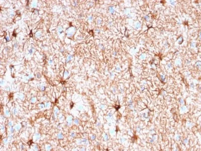
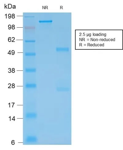
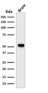
This MAb recognizes a protein of ~50 kDa which is identified as Glial Fibrillary Acidic Protein (GFAP). It shows no cross-reaction with other intermediate filament proteins. GFAP is specifically found in astroglia. GFAP is a very popular marker for localizing benign astrocyte and neoplastic cells of glial origin in the central nervous system. Antibody to GFAP is useful in differentiating primary gliomas from metastatic lesions in the brain and for documenting astrocytic differentiation in tumors outside the CNS.Primary antibodies are available purified, or with a selection of fluorescent CF® Dyes and other labels. CF® Dyes offer exceptional brightness and photostability. Note: Conjugates of blue fluorescent dyes like CF®405S and CF®405M are not recommended for detecting low abundance targets, because blue dyes have lower fluorescence and can give higher non-specific background than other dye colors.Synonyms:
Astrocyte or Intermediate Filament Protein, Glial Fibrillary Acidic Protein (GFAP)UNSPSC:
41116161UNSPSC Description:
Primary and secondary antibodies for multiple methodology immunostaining detection applicationGene Name:
GFAPGene ID:
2670NCBI Gene ID:
514227UniProt:
P14136Cellular Locus:
CytoskeletonHost:
MouseSpecies Reactivity:
Chicken, Cow, Human, Mouse, Pig, Rabbit, RatImmunogen:
Recombinant full-length human GFAP proteinTarget Antigen:
GFAPClonality:
Recombinant MonoclonalIsotype:
IgG1Clone:
rASTRO/789Conjugation:
CF640RSource:
AnimalApplications:
Flow, intracellular (verified) | IHC, FFPE (verified) | WB (verified)Validated Applications:
FC, IHC, FFPE, WBField of Research:
NeurosciencePositive Control:
Brain or AstrocytomaConcentration:
0.1 mg/mLBuffer:
PBS, 0.1% BSA, 0.05% azideMolecular Weight:
~50 kDaAdditionnal Information:
Higher concentration may be required for direct detection using primary antibody conjugates than for indirect detection with secondary antibody|Immunohistology (formalin): 0.25-0.5 ug/mL for 30 minutes at RT|Staining of formalin-fixed tissues requires boiling tissue sections in 10 mM citrate buffer, pH 6.0, for 10-20 minutes followed by cooling at RT for 20 minutes|Western blotting 0.5-1 ug/mL|Optimal dilution for a specific application should be determined by userShipping Conditions:
Room temperatureStorage Conditions:
4°C; Protect from light; Stable at room temperature or 37°C (98°F) for 7 days.Shelf Life:
2 yearsCAS Number:
9007-83-4
DATASHEET Document
View DocumentMSDS Document
View Document