Anti-TCL1 (T-Cell Marker) (TCL1/2747R)
CAT:
37-BNUB2747-500
Size:
500 µL
Price:
Ask
- Availability: 24/48H Stock Items & 2 to 6 Weeks non Stock Items.
- Dry Ice Shipment: No


Anti-TCL1 (T-Cell Marker) (TCL1/2747R)
Description:
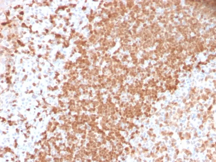
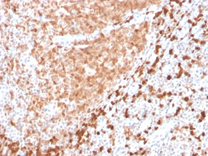
T-cell leukemia/Lymphoma Protein 1A (TCL-1A), also known as p14TCL1 is a product of the TCL1 gene that is involved in T-cell prolymphocytic leukemia (TPLL). T-PLL is a rare form of mature T-cell leukemia, which is consistently associated with chromosomal rearrangements characterized by the juxtaposition of the TCRA locus on chromosome 14q11 and the TCL1A gene on 14q32.13. TCL1 is overexpressed in Burkitt lymphoma, the majority of AIDS-related non-Hodgkin lymphoma-designated immunoblastic plasmacytoid lymphoma, lymphoblastic lymphoma, chronic lymphocytic leukemia, mantle cell lymphoma, follicular lymphoma, diffuse large B-cell lymphoma, and primary cutaneous B-cell lymphoma.Primary antibodies are available purified, or with a selection of fluorescent CF® Dyes and other labels. CF® Dyes offer exceptional brightness and photostability. Note: Conjugates of blue fluorescent dyes like CF®405S and CF®405M are not recommended for detecting low abundance targets, because blue dyes have lower fluorescence and can give higher non-specific background than other dye colors.Synonyms:
TCL1A; Lymphoma/leukemia, T-cell; Oncogene TCL1; P14 TCL1; T cell leukemia 1; T cell lymphoma 1; T cell lymphoma 1A;T-cell leukemia/lymphoma 1A; TCL1; TCL1AUNSPSC:
41116161UNSPSC Description:
Primary and secondary antibodies for multiple methodology immunostaining detection applicationGene Name:
TCL1AGene ID:
10232NCBI Gene ID:
2484UniProt:
P56279Cellular Locus:
Nucleus & cytoplasmHost:
RabbitSpecies Reactivity:
HumanImmunogen:
Recombinant human TCL1 protein fragment (around aa 2-109) (exact sequence is proprietary)Target Antigen:
TCL1Clonality:
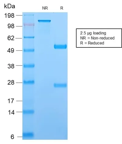
Recombinant MonoclonalIsotype:
IgGClone:
TCL1/2747RConjugation:
Purified, with BSASource:
AnimalApplications:
IHC, FFPE (verified)Validated Applications:
IHC, FFPEField of Research:
Cancer, ImmunologyPositive Control:
Ramos or Raji cells. Tonsil or T-cell Lymphoma.Concentration:
0.2 mg/mLBuffer:
PBS, 0.05% BSA, 0.05% azideMolecular Weight:
14 kDaAdditionnal Information:
Higher concentration may be required for direct detection using primary antibody conjugates than for indirect detection with secondary antibody|Immunohistology (formalin): 1-2 ug/mL for 30 minutes at RT|Staining of formalin-fixed tissues requires boiling tissue sections in 10 mM citrate buffer, pH 6.0, for 10-20 minutes followed by cooling at RT for 20 minutes|Optimal dilution for a specific application should be determined by userShipping Conditions:
Room temperatureStorage Conditions:
4°C; Stable at room temperature or 37°C (98°F) for 7 days.Shelf Life:
2 yearsCAS Number:
9007-83-4
DATASHEET Document
View DocumentMSDS Document
View Document