
Anti-Villin (GI-Mucosal & Urogenital Brush Border Marker) (rVIL1/1325)
CAT:
37-BNUB2279-500
Size:
500 µL
Price:
Ask
- Availability: 24/48H Stock Items & 2 to 6 Weeks non Stock Items.
- Dry Ice Shipment: No


Anti-Villin (GI-Mucosal & Urogenital Brush Border Marker) (rVIL1/1325)
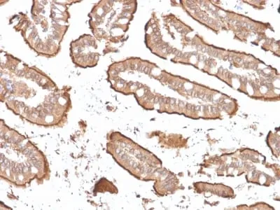
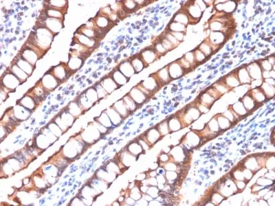
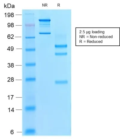
- Description: This antibody recognizes a protein of 95 kDa, which is identified as villin. It is a major constituent in the microvilli, which compose the brush border of epithelial cells forming absorptive surfaces of the intestinal and renal proximal tubular epithelia. Anti-Villin labels the brush border area in the gastrointestinal mucosal epithelium and urogenital tract. Among neoplasms, villin is predominantly expressed in tumors of colorectal origin. Antibody to villin is useful in identifying malignant cells from primary and metastatic colorectal carcinomas. This antibody also labels Merkel cells of the skin.Primary antibodies are available purified, or with a selection of fluorescent CF® Dyes and other labels. CF® Dyes offer exceptional brightness and photostability. Note: Conjugates of blue fluorescent dyes like CF®405S and CF®405M are not recommended for detecting low abundance targets, because blue dyes have lower fluorescence and can give higher non-specific background than other dye colors.
- Synonyms: VIL1; Villin-1; Villin1
- CAS Number: 9007-83-4
- UNSPSC: 41116161
- UNSPSC Description: Primary and secondary antibodies for multiple methodology immunostaining detection application
- Gene Name: VIL1
- Gene ID: 7429
- NCBI Gene ID: 654595
- UniProt: P09327
- Cellular Locus: Cytoskeleton
- Host: Mouse
- Species Reactivity: Human
- Immunogen: Recombinant human Villin fragment of 133 amino acid residues (aa179-311)
- Target Antigen: Villin
- Clonality: Recombinant Monoclonal
- Isotype: IgG1 κ
- Clone: rVIL1/1325
- Conjugation: Purified, with BSA
- Disease: Tumor
- Source: Animal
- Applications: IHC, FFPE (verified)
- Validated Applications: IHC, FFPE
- Field of Research: Cancer
- Positive Control: A549, HepG2 and HCT116 cells. Colon or Rectum.
- Concentration: 0.2 mg/mL
- Buffer: PBS, 0.05% BSA, 0.05% azide
- Molecular Weight: 93 kDa
- Additionnal Information: Higher concentration may be required for direct detection using primary antibody conjugates than for indirect detection with secondary antibody|Immunohistology (formalin): 1-2 ug/mL for 30 minutes at RT|Staining of formalin-fixed tissues requires boiling tissue sections in 10 mM citrate buffer, pH 6.0, for 10-20 minutes followed by cooling at RT for 20 minutes|Optimal dilution for a specific application should be determined by user
- Shipping Conditions: Room temperature
- Storage Conditions: 4°C; Stable at room temperature or 37°C (98°F) for 7 days.
- Shelf Life: 2 years