Anti-p63 (Squamous, Basal and Myoepithelial Cell Marker) (TP63/1786)
CAT:
37-BNUM1786-50
Size:
50 µL
Price:
Ask
- Availability: 24/48H Stock Items & 2 to 6 Weeks non Stock Items.
- Dry Ice Shipment: No


Anti-p63 (Squamous, Basal and Myoepithelial Cell Marker) (TP63/1786)
Description:
p63 is a homolog of the tumor suppressor p53. It is identified in basal cells in the epithelial layers of a variety of tissues, including epidermis, cervix, urothelium, breast and prostate. p63 was detected in nuclei of the basal epithelium in normal prostate glands; however, it was not expressed in malignant tumors of the prostate. As a result, p63 has been reported as a useful marker for differentiating benign from malignant lesions in the prostate, particularly when used in combination with markers of high molecular weight cytokeratins and the prostate-specific marker AMACR (P504S). p63 has also been shown to be a sensitive marker for lung squamous cell carcinomas (SqCC), with a sensitivity of ~90%. Specificity for lung SqCC, vs. lung adenocarcinoma (LADC), is approximately 80%. In breast tissue, p63 has been identified in myoepithelial cells of normal ducts._x000D_ _x000D_ Primary antibodies are available purified, or with a selection of fluorescent CF® Dyes and other labels. CF® Dyes offer exceptional brightness and photostability. Note: Conjugates of blue fluorescent dyes like CF®405S and CF®405M are not recommended for detecting low abundance targets, because blue dyes have lower fluorescence and can give higher non-specific background than other dye colors._x000D_ _x000D_Synonyms:
Amplified in squamous cell carcinoma (AIS); Chronic ulcerative stomatitis protein (CUSP); EEC3; Keratinocyte transcription factor KET; LMS; NBP; p40; P51/P63; p53 like transcription factor; p53-related protein p63; RHS; SHFM4; TAp63alpha; TP53CP; TP53L; TP63; TP73; TP73L; Transformation-related protein 63; Trp53rp1; Trp6; 3; Tumor protein 63; Tumor protein p53-like; tumor protein p73-likeUNSPSC:
41116161UNSPSC Description:
Primary and secondary antibodies for multiple methodology immunostaining detection applicationGene Name:
TP63Gene ID:
8626NCBI Gene ID:
137569UniProt:
Q9H3D4Cellular Locus:
NucleusHost:
MouseSpecies Reactivity:
HumanImmunogen:
Recombinant human p63 protein fragment (aa3-106) (exact sequence is proprietary)Target Antigen:
p63Clonality:
MonoclonalIsotype:
IgG2b κClone:
TP63/1786Conjugation:
Purified, BSA-freeDisease:
TumorSource:
AnimalApplications:
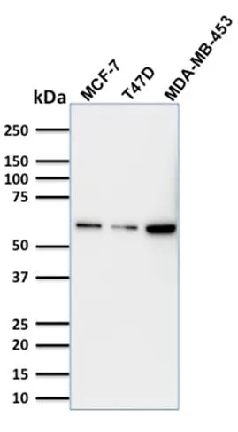
WB (verified)Validated Applications:
WBField of Research:
Cancer, Cell cycle, Tumor suppressorsPositive Control:
HEK293 cells or Prostate Carcinoma or Lung or bladder squamous cell carcinomaConcentration:
1 mg/mLBuffer:
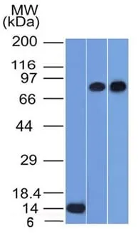
PBS, no BSA, no azideMolecular Weight:
63 kDaAdditionnal Information:
Higher concentration may be required for direct detection using primary antibody conjugates than for indirect detection with secondary antibody|ELISA: For coating order antibody without BSA|Western blotting 1-2 ug/mL|Optimal dilution for a specific application should be determined by userShipping Conditions:
Room temperatureStorage Conditions:
-35°C to -5°C ; Stable at room temperature or 37°C (98°F) for 7 days.Shelf Life:
2 yearsCAS Number:
9007-83-4
DATASHEET Document
View DocumentMSDS Document
View Document