Anti-Ferritin, Light Chain (FTL) (Microglia Marker) (FTL/1386), CF640R conjugate
CAT:
37-BNC401386-500
Size:
500 µL
Price:
Ask
- Availability: 24/48H Stock Items & 2 to 6 Weeks non Stock Items.
- Dry Ice Shipment: No


Anti-Ferritin, Light Chain (FTL) (Microglia Marker) (FTL/1386), CF640R conjugate
Description:
Mammalian ferritins consist of 24 subunits made up of 2 types of polypeptide chains, ferritin heavy chain and ferritin light chain. Ferritin heavy chains catalyze the first step in iron storage, the oxidation of Fe (II), whereas ferritin light chains promote the nucleation of ferrihydrite, enabling storage of Fe (III). Light chain ferritin is involved in cataracts by at least two mechanisms, hereditary hyperferritinemia cataract syndrome, in which light chain ferritin is overexpressed, and oxidative stress, an important factor in the development of ageing-related cataracts.Primary antibodies are available purified, or with a selection of fluorescent CF® Dyes and other labels. CF® Dyes offer exceptional brightness and photostability. Note: Conjugates of blue fluorescent dyes like CF®405S and CF®405M are not recommended for detecting low abundance targets, because blue dyes have lower fluorescence and can give higher non-specific background than other dye colors.Synonyms:
Ferritin L chain; Ferritin L subunit; Ferritin light chain; Ferritin light polypeptide; FTL; LFTD; NBIA3UNSPSC:
41116161UNSPSC Description:
Primary and secondary antibodies for multiple methodology immunostaining detection applicationGene Name:
FTLGene ID:
2512NCBI Gene ID:
433670UniProt:
P02792Cellular Locus:
CytoplasmicHost:
MouseSpecies Reactivity:
HumanImmunogen:
Recombinant human FTL protein fragment (aa 38-165) (exact sequence is proprietary)Target Antigen:
Ferritin, Light ChainClonality:
MonoclonalIsotype:
IgG2bClone:
FTL/1386Conjugation:
CF640RSource:
AnimalApplications:
IHC, FFPE (verified) | WB (verified)Validated Applications:
IHC, FFPE, WBField of Research:
MetabolismPositive Control:
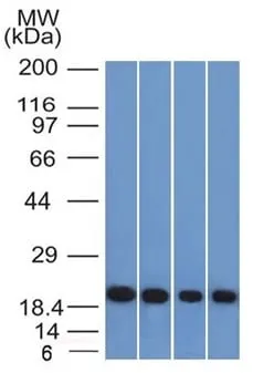
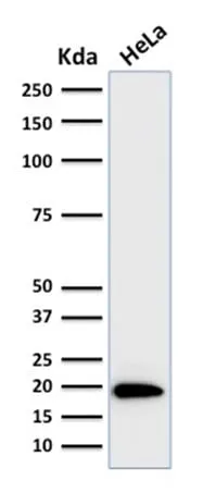
HepG2, HeLa, HL-60 or 293T cells. Pancreas, Liver, Cerebellum or Testis.Concentration:
0.1 mg/mLBuffer:
PBS, 0.1% BSA, 0.05% azideMolecular Weight:
19-25 kDaAdditionnal Information:
Higher concentration may be required for direct detection using primary antibody conjugates than for indirect detection with secondary antibody|Immunohistology (formalin) 0.1-0.2 ug/mL|Flow cytometry 0.1-0.2ug/million cells Immunofluorescence 0.1-0.2ug/ml|Western blotting 0.1-0.2ug/ml|Staining of formalin-fixed tissues requires boiling tissue sections in 10 mM citrate buffer, pH 6.0, for 10-20 min followed by cooling at RT for 20 min|Optimal dilution for a specific application should be determined by userShipping Conditions:
Room temperatureStorage Conditions:
4°C; Protect from light; Stable at room temperature or 37°C (98°F) for 7 days.Shelf Life:
2 yearsCAS Number:
9007-83-4
DATASHEET Document
View DocumentMSDS Document
View Document