
Anti-TRIM29(TRIM29/1041)
CAT:
37-BNUB1041-500
Size:
500 µL
Price:
Ask
- Availability: 24/48H Stock Items & 2 to 6 Weeks non Stock Items.
- Dry Ice Shipment: No


Anti-TRIM29(TRIM29/1041)
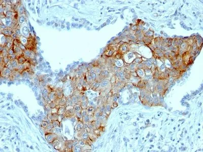
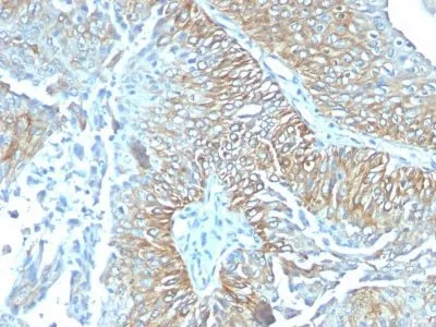
- Description: This antibody recognizes a 66 kDa protein, which is identified as Tripartite motif-containing protein 29 (TRIM29). It interacts with the intermediate filament protein vimentin, a substrate for the PKC family of protein kinases, and with hPKCI-1, an inhibitor of the PKCs. TRIM29 protein contains both zinc finger and leucine zipper motifs, suggesting that the it may form homodimers and possibly associate with DNA. High expression of TRIM29 has been reported in gastric cancer and pancreatic cancer, and correlates with enhanced tumor growth and lymph node metastasis. TRIM29 is also able to distinguish lung squamous cell carcinoma from lung adenocarcinoma with ~90% positive accuracy, when used in a panel with TTF-1, p63, CK5/6, and Napsin-A antibodies.Primary antibodies are available purified, or with a selection of fluorescent CF® Dyes and other labels. CF® Dyes offer exceptional brightness and photostability. Note: Conjugates of blue fluorescent dyes like CF®405S and CF®405M are not recommended for detecting low abundance targets, because blue dyes have lower fluorescence and can give higher non-specific background than other dye colors.
- Synonyms: Ataxia telangiectasia group D complementing gene (ATDC); Tripartite motif-containing protein 29 (TRIM29);
- CAS Number: 9007-83-4
- UNSPSC: 41116161
- UNSPSC Description: Primary and secondary antibodies for multiple methodology immunostaining detection application
- Gene Name: TRIM29
- Gene ID: 23650
- NCBI Gene ID: 504115
- UniProt: Q14134
- Cellular Locus: Cytoplasmic|Cytoskeleton|Nucleus
- Host: Mouse
- Species Reactivity: Human
- Immunogen: Recombinant fragment (126 Amino acid residues between aa 1-200) of human TRIM29 protein
- Target Antigen: TRIM29
- Clonality: Monoclonal
- Isotype: IgG2a κ
- Clone: TRIM29/1041
- Conjugation: Purified, with BSA
- Disease: Tumor
- Source: Animal
- Applications: IHC, FFPE (verified)
- Validated Applications: IHC, FFPE
- Field of Research: Cancer, Cytoskeleton, Signal transduction
- Positive Control: A431 cells. Tonsil or Squamous cell carcinoma.
- Concentration: 0.2 mg/mL
- Buffer: PBS, 0.05% BSA, 0.05% azide
- Molecular Weight: 66 kDa
- Additionnal Information: Higher concentration may be required for direct detection using primary antibody conjugates than for indirect detection with secondary antibody|Immunofluorescence: 0.5-1 ug/mL|Immunohistology formalin-fixed 0.5-1 ug/mL|Staining of formalin-fixed tissues requires boiling tissue sections in 10 mM citrate buffer, pH 6.0, for 10-20 min followed by cooling at RT for 20 minutes|Flow Cytometry 0.5-1 ug/million cells/0.1 mL|Optimal dilution for a specific application should be determined by user
- Shipping Conditions: Room temperature
- Storage Conditions: 4°C; Stable at room temperature or 37°C (98°F) for 7 days.
- Shelf Life: 2 years