
Anti-TGF-beta(1D11.16.8)
CAT:
37-BNUB0977-100
Size:
100 µL
Price:
Ask
- Availability: 24/48H Stock Items & 2 to 6 Weeks non Stock Items.
- Dry Ice Shipment: No


Anti-TGF-beta(1D11.16.8)
- Description: This MAb recognizes TGF beta 1, 2 and 3. Three TGF-betas have been identified in mammals. TGF-beta1, TGF-beta2 and TGF-beta3 are each synthesized as precursor proteins that are very similar in that each is cleaved to yield a 112 amino acid polypeptide that remains associated with the latent portion of the molecules. Biologically active TGF-beta requires dimerization of the monomers (usually homodimers) and release of the latent peptide portion. Overall, the mature region of the TGF-beta3 protein has approximately 80% identity to the mature region of both TGF-beta1 and TGF-beta2. However, the NH2 terminals or precursor regions of their molecules share only 27% sequence identity. TGF-beta's inhibit the growth of epithelial cells and stimulate the growth of mesenchymal cells.Primary antibodies are available purified, or with a selection of fluorescent CF® Dyes and other labels. CF® Dyes offer exceptional brightness and photostability. Note: Conjugates of blue fluorescent dyes like CF®405S and CF®405M are not recommended for detecting low abundance targets, because blue dyes have lower fluorescence and can give higher non-specific background than other dye colors.
- Synonyms: GP40; Leu9; p41; T-cell leukemia antigen; T-cell surface antigen Leu-9; Tp40; TP41
- CAS Number: 9007-83-4
- UNSPSC: 41116161
- UNSPSC Description: Primary and secondary antibodies for multiple methodology immunostaining detection application
- Gene Name: TGFB1
- Gene ID: 7040 (beta1), 7042 (beta2) & 7043 (beta3)
- NCBI Gene ID: 645227
- UniProt: P01137 (beta1), P10600 (beta2); P61812 (beta3)
- Cellular Locus: Secreted (extracellular)
- Host: Mouse
- Species Reactivity: Cow, Dog, Hamster, Human, Monkey, Mouse, Rat
- Immunogen: Bovine TGF-beta2 protein
- Target Antigen: TGF-Beta
- Clonality: Monoclonal
- Isotype: IgG1 κ
- Clone: 1D11.16.8
- Conjugation: Purified, with BSA
- Source: Animal
- Applications: Functional studies (published) | IHC, FFPE (published)
- Validated Applications: IHC, FFPE
- Field of Research: Signal transduction
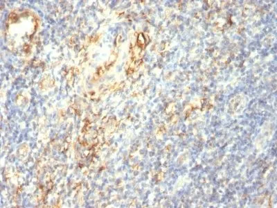
- Positive Control: Salivary gland carcinoma
- Concentration: 0.2 mg/mL
- Buffer: PBS, 0.05% BSA, 0.05% azide
- Molecular Weight: ~13 kDa
- Additionnal Information: Higher concentration may be required for direct detection using primary antibody conjugates than for indirect detection with secondary antibody|Cytokine neutralization or functional assay, order Ab without BSA/azide|Optimal dilution for a specific application should be determined by user
- References & Citations: Note: References for this clone sold by other suppliers may be listed for expected applications. Biol Reproduct (1992) 46: 561-572. (IHC, FFPE) PLOS ONE 10(7): e0132786. (functional studies)
- Shipping Conditions: Room temperature
- Storage Conditions: 4°C; Stable at room temperature or 37°C (98°F) for 7 days.
- Shelf Life: 2 years