
Anti-Tenascin(T2H5)
- Availability: 24/48H Stock Items & 2 to 6 Weeks non Stock Items.
- Dry Ice Shipment: No


Anti-Tenascin(T2H5)
Description:
In Western blotting, this antibody reacts with two bands of ~MW of 210 kDa and 300 kDa, identified as two isoforms of Tenascin C. Specificity of this MAb is validated by sequential immunoprecipitation with a PAb against Tenascin C. Tenascin C is a multifunctional, disulfide-linkedhexameric extracellular matrix glycoprotein expressed in association with mesenchymal epithelial interactions during development and in the neo-vasculature and stroma of undifferentiated tumors. In adults, it is restricted to certain epithelial-stromal interfaces and increases markedly in hyper-proliferative diseases and in stroma of many neoplasms, including gliomas, breast, squamous and lung carcinomas.Primary antibodies are available purified, or with a selection of fluorescent CF® Dyes and other labels. CF® Dyes offer exceptional brightness and photostability. Note: Conjugates of blue fluorescent dyes like CF®405S and CF®405M are not recommended for detecting low abundance targets, because blue dyes have lower fluorescence and can give higher non-specific background than other dye colors.Synonyms:
Cytotactin; Glioma-associated-extracellular matrix antigen; GMEM; GP 150-225; Hexabrachion (HXB); JI; Myotendinous antigen; Neuronectin; TNCUNSPSC:
41116161UNSPSC Description:
Primary and secondary antibodies for multiple methodology immunostaining detection applicationGene Name:
TNCGene ID:
3371NCBI Gene ID:
143250UniProt:
P24821Cellular Locus:
Extracellular matrixHost:
MouseSpecies Reactivity:
HumanImmunogen:
Human breast carcinomaTarget Antigen:
TenascinClonality:
MonoclonalIsotype:
IgG1 κClone:
T2H5Conjugation:
Purified, with BSASource:
AnimalApplications:
IHC, FFPE (verified)Validated Applications:
IHC, FFPEField of Research:
CancerPositive Control:
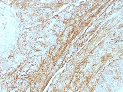
Extracellular matrix in tonsil and blood vessels. Stroma of many tumors such as breast, squamous cell, and lung carcinomas. Staining of normal fibroblasts serves as internal positive controlConcentration:
0.2 mg/mLBuffer:
PBS, 0.05% BSA, 0.05% azideMolecular Weight:
210 kDa and 300 kDaAdditionnal Information:
Higher concentration may be required for direct detection using primary antibody conjugates than for indirect detection with secondary antibody|Immunofluorescence: 0.5-1 ug/mL|Does not react with rat; others not known|Immunohistology formalin-fixed 2-4 ug/mL|Staining of formalin-fixed tissues requires boiling tissue sections in 10 mM Tris with 1 mM EDTA, pH 9.0, for 10-20 min followed by cooling at RT for 20 minutes|Flow Cytometry 0.5-1 ug/million cells/0.1 mL|Optimal dilution for a specific application should be determined by userShipping Conditions:
Room temperatureStorage Conditions:
4°C; Stable at room temperature or 37°C (98°F) for 7 days.Shelf Life:
2 yearsCAS Number:
9007-83-4
DATASHEET Document
View DocumentMSDS Document
View Document