Anti-Pmel17 / gp100 / SILV(NKI-beteb)
CAT:
37-BNUM0226-50
Size:
50 µL
Price:
Ask
- Availability: 24/48H Stock Items & 2 to 6 Weeks non Stock Items.
- Dry Ice Shipment: No


Anti-Pmel17 / gp100 / SILV(NKI-beteb)
Description:
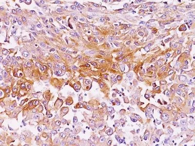
By immunohistochemistry, this antibody specifically recognizes a protein in melanocytes and melanomas. It reacts with junctional and blue nevus cells and variably with fetal and neonatal melanocytes. Intradermal nevi, normal adult melanocytes, and non-melanocytic cells are negative. It does not stain tumor cells of epithelial, lymphoid, glial, or mesenchymal origin. This Mab labels formalin-fixed, paraffin-embedded melanomas and other tumors showing melanocytic differentiation.Primary antibodies are available purified, or with a selection of fluorescent CF® Dyes and other labels. CF® Dyes offer exceptional brightness and photostability. Note: Conjugates of blue fluorescent dyes like CF®405S and CF®405M are not recommended for detecting low abundance targets, because blue dyes have lower fluorescence and can give higher non-specific background than other dye colors.Synonyms:
95kDa melanocyte-specific secreted glycoprotein, M-beta, Melanocyte lineage specific antigen GP100, Melanocyte protein Pmel 17, Melanoma associated ME20 antigen, Melanosomal matrix protein17, p100, p26, PMEL17, Premelanosome protein, Secreted melanoma-associated ME20 antigen, SILV, Silver homologUNSPSC:
41116161UNSPSC Description:
Primary and secondary antibodies for multiple methodology immunostaining detection applicationGene Name:
PMELGene ID:
6490NCBI Gene ID:
95972UniProt:
P40967Cellular Locus:
Endoplasmic reticulum|Golgi apparatusHost:
MouseSpecies Reactivity:
Horse, HumanImmunogen:
Membranes from a human melanoma metastasisTarget Antigen:
gp100 | Pmel17 | SILVClonality:
MonoclonalIsotype:
IgG2b κClone:
NKI-betebConjugation:
Purified, BSA-freeDisease:
TumorSource:
AnimalApplications:
Flow, intracellular (published) | IF (published) | IHC, frozen (published) | ELISA (published) | IHC, FFPE (verified) | IP (published)Validated Applications:
FC, IF, IHC, ELISA, IHC, FFPE, IPField of Research:
CancerPositive Control:
SK-MEL-28 cells or MelanomaConcentration:
1 mg/mLBuffer:
PBS, no BSA, no azideMolecular Weight:
90-100 kDaAdditionnal Information:
Immunohistology formalin-fixed 1-2 ug/mL|Staining of formalin-fixed tissues requires boiling tissue sections in 10 mM citrate buffer, pH 6.0, for 10-20 min followed by cooling at RT for 20 minutes|Flow Cytometry 0.5-1 ug/million cells/0.1 mL|Immunofluorescence 1-2 ug/mL|Does not react with rat, others not tested|Optimal dilution for a specific application should be determined by userReferences & Citations:
Note: References for this clone sold by other suppliers may be listed for expected applications. Am J Pathol (1988) 130(1): 179. (mAb characterization; IHC, FFPE; IF; ELISA; IP; WB) Am J Pathol (1993) 143(6): 1579. (IHC, frozen; IF) J Biol Chem (2004) 279(27): 28330-28338. (epitope mapping; IP; reports that NKI-beteb does not work well for WB) Exp Dermatol (2010) 19(8): e282-e285. (IF; Flow, intracellular)Shipping Conditions:
Room temperatureStorage Conditions:
-35°C to -5°C ; Stable at room temperature or 37°C (98°F) for 7 days.Shelf Life:
2 yearsCAS Number:
9007-83-4
DATASHEET Document
View DocumentMSDS Document
View Document