Anti-pgp9.5 (UCHL-1)(31A3)
CAT:
37-BNUM0046-50
Size:
50 µL
Price:
Ask
- Availability: 24/48H Stock Items & 2 to 6 Weeks non Stock Items.
- Dry Ice Shipment: No


Anti-pgp9.5 (UCHL-1)(31A3)
Description:
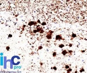
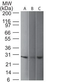
This MAb reacts with a protein of 20-30 kDa, identified as PGP9.5, also known as ubiquitin carboxyl-terminal hydrolase-1 (UchL1). Initially, PGP9.5 expression in normal tissues was reported in neurons and neuroendocrine cells but later it was found in distal renal tubular epithelium, spermatogonia, Leydig cells, oocytes, melanocytes, prostatic secretory epithelium, ejaculatory duct cells, epididymis, mammary epithelial cells, Merkel cells, and dermal fibroblasts. Furthermore, immunostaining for PGP9.5 has been shown in a wide variety of mesenchymal neoplasms as well. A mutation in PGP9.5 gene is believed to cause a form of Parkinson's disease.Primary antibodies are available purified, or with a selection of fluorescent CF® Dyes and other labels. CF® Dyes offer exceptional brightness and photostability. Note: Conjugates of blue fluorescent dyes like CF®405S and CF®405M are not recommended for detecting low abundance targets, because blue dyes have lower fluorescence and can give higher non-specific background than other dye colors.Synonyms:
Gracile Axonal Dystrophy|Neuron Cytoplasmic Protein 9.5|Park5|Parkinson Disease 5|PGP95|Protein Gene Product 9.5|Ubiquitin Carboxyl-terminal Esterase L1|Ubiquitin Carboxyl-terminal Hydrolase Isozyme L1|Ubiquitin Thioesterase L1|Ubiquitin Thiolesterase L1UNSPSC:
41116161UNSPSC Description:
Primary and secondary antibodies for multiple methodology immunostaining detection applicationGene Name:
UCHL1Gene ID:
7345NCBI Gene ID:
518731UniProt:
P09936Cellular Locus:
Cytoplasmic|Endoplasmic reticulumHost:
MouseSpecies Reactivity:
Cow, Human, Mouse, Pig, RatImmunogen:
Native UchL1 (PGP9.5) protein from brainTarget Antigen:
PGP9.5 | UchL1Clonality:
MonoclonalIsotype:
IgG1 κClone:
31A3Conjugation:
Purified, BSA-freeSource:
AnimalApplications:
Flow, intracellular (verified) | IF (verified) | IHC, FFPE (verified) | WB (verified)Validated Applications:
FC, IF, IHC, FFPE, WBField of Research:
Metabolism, NeurosciencePositive Control:
CerebellumConcentration:
1 mg/mLBuffer:
PBS, no BSA, no azideMolecular Weight:
20-30 kDaAdditionnal Information:
Higher concentration may be required for direct detection using primary antibody conjugates than for indirect detection with secondary antibody|Immunohistology formalin-fixed 0.5-1 ug/mL|Staining of formalin-fixed tissues requires boiling tissue sections in 10 mM citrate buffer, pH 6.0, for 10-20 min followed by cooling at RT for 20 minutes|Western blotting 0.5-1 ug/mL|Optimal dilution for a specific application should be determined by userShipping Conditions:
Room temperatureStorage Conditions:
-35°C to -5°C ; Stable at room temperature or 37°C (98°F) for 7 days.Shelf Life:
2 yearsCAS Number:
9007-83-4
DATASHEET Document
View DocumentMSDS Document
View Document