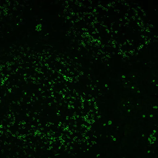
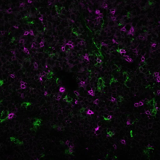
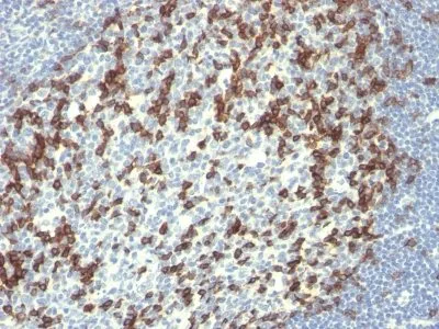
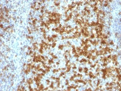
Anti-PD1 / PDCD1 / CD279(PDCD1/922), CF640R conjugate
CAT:
37-BNC400922-500
Size:
500 µL
Price:
Ask
- Availability: 24/48H Stock Items & 2 to 6 Weeks non Stock Items.
- Dry Ice Shipment: No


Anti-PD1 / PDCD1 / CD279(PDCD1/922), CF640R conjugate
Description:
PD-1 (also known as Programmed Death 1, CD279, and PDCD1) is a negative regulatory T cell surface receptor. PD-1 is a Type I transmembrane protein expressed on the plasma membrane of T-cells. It is a T-cell checkpoint protein, involved in preventing autoimmunity. Because binding of PD-1 by its ligands PD-L1 or PD-L2 has a suppressive effect on the immune system, inhibition of the PD-1/PD-L1 pathway is a major strategy of immune-oncology therapies. PD-L1 is upregulated in certain cancers, and blocking PD-1 or PD-L1 can inhibit cancer growth._x000D_ _x000D_ Primary antibodies are available purified, or with a selection of fluorescent CF® Dyes and other labels. CF® Dyes offer exceptional brightness and photostability. Note: Conjugates of blue fluorescent dyes like CF®405S and CF®405M are not recommended for detecting low abundance targets, because blue dyes have lower fluorescence and can give higher non-specific background than other dye colors._x000D_ _x000D_Synonyms:
CD279; hPD-1; hSLE1; PD1; PDCD1; Programmed Cell Death Protein 1; Protein PD-1; SLEB2; Systemic lupus erythematosus susceptibility 2UNSPSC:
41116161UNSPSC Description:
Primary and secondary antibodies for multiple methodology immunostaining detection applicationGene Name:
PDCD1Gene ID:
5133NCBI Gene ID:
158297UniProt:
Q15116Cellular Locus:
Plasma membraneHost:
MouseSpecies Reactivity:
HumanImmunogen:
Recombinant full-length human PDCD1 proteinTarget Antigen:
CD279 | PD1 | PDCD1Clonality:
MonoclonalIsotype:
IgG1 κClone:
PDCD1/922Conjugation:
CF640RDisease:
TumorSource:
AnimalApplications:
Flow (verified) | IF (verified) | IHC, FFPE (verified)Validated Applications:
FC, IF, IHC, FFPEField of Research:
Apoptosis, Cancer, Cancer immunotherapy/immune checkpointPositive Control:
TY cells or Tonsil.Concentration:
0.1 mg/mLBuffer:
PBS, 0.1% BSA, 0.05% azideMolecular Weight:
55 kDaAdditionnal Information:
Higher concentration may be required for direct detection using primary antibody conjugates than for indirect detection with secondary antibody|Immunofluorescence: 1-2 ug/mL|Immunohistology formalin-fixed 0.5-1 ug/mL|Staining of formalin-fixed tissues requires boiling tissue sections in 10 mM Tris with 1 mM EDTA, pH 9.0, for 10-20 min followed by cooling at RT for 20 minutes|Flow Cytometry 0.5-1 ug/million cells/0.1 mL|Optimal dilution for a specific application should be determined by userShipping Conditions:
Room temperatureStorage Conditions:
4°C; Protect from light; Stable at room temperature or 37°C (98°F) for 7 days.Shelf Life:
2 yearsCAS Number:
9007-83-4
DATASHEET Document
View DocumentMSDS Document
View Document