
Anti-PCNA(PCNA/694), CF405S conjugate
- Availability: 24/48H Stock Items & 2 to 6 Weeks non Stock Items.
- Dry Ice Shipment: No


Anti-PCNA(PCNA/694), CF405S conjugate
Description:
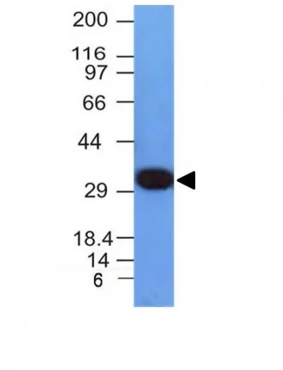
Recognizes a non-histone protein of 36 kDa, which is identified as proliferating cell nuclear antigen (PCNA). It is also known as cyclin or polymerase delta auxiliary protein. Elevated expression of PCNA/cyclin has been shown in the nucleus during late G1 phase immediately before the onset of DNA synthesis, becoming maximal during S-phase and declining during G2 and M phases. This MAb is excellent for multiple applications.Primary antibodies are available purified, or with a selection of fluorescent CF® Dyes and other labels. CF® Dyes offer exceptional brightness and photostability. Note: Conjugates of blue fluorescent dyes like CF®405S and CF®405M are not recommended for detecting low abundance targets, because blue dyes have lower fluorescence and can give higher non-specific background than other dye colors.Synonyms:
Cyclin; DNA polymerase delta auxiliary protein; Mutagen-sensitive 209 protein; PCNAR; Polymerase delta accessory proteinUNSPSC:
41116161UNSPSC Description:
Primary and secondary antibodies for multiple methodology immunostaining detection applicationGene Name:
PCNAGene ID:
5111NCBI Gene ID:
147433 & 728886UniProt:
P12004Cellular Locus:
Nucleus & cytoplasmHost:
MouseSpecies Reactivity:
HumanImmunogen:
Recombinant full length human PCNA proteinTarget Antigen:
PCNAClonality:
MonoclonalIsotype:
IgG2a κClone:
PCNA/694Conjugation:
CF405SSource:
AnimalApplications:
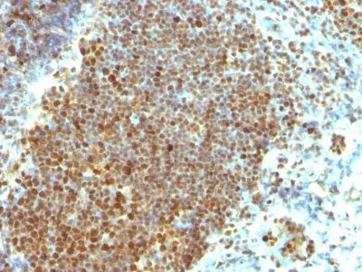
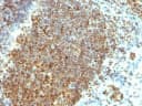
Flow, intracellular (verified) | IHC, FFPE (verified) | WB (verified)Validated Applications:
FC, IHC, FFPE, WBField of Research:
Cell cyclePositive Control:
Tonsil or reactive lymph nodeConcentration:
0.1 mg/mLBuffer:
PBS, 0.1% BSA, 0.05% azideMolecular Weight:
36 kDaAdditionnal Information:
Higher concentration may be required for direct detection using primary antibody conjugates than for indirect detection with secondary antibody|Immunofluorescence: 0.5-1 ug/mL|Immunohistology formalin-fixed 0.25-0.5 ug/mL|Staining of formalin-fixed tissues requires boiling tissue sections in 10 mM citrate buffer, pH 6.0, for 10-20 min followed by cooling at RT for 20 minutes|Flow Cytometry 0.5-1 ug/million cells/0.1 mL|Western blotting 0.5-1 ug/mL|Predicted to show broad species reactivity|Optimal dilution for a specific application should be determined by userShipping Conditions:
Room temperatureStorage Conditions:
4°C; Protect from light; Stable at room temperature or 37°C (98°F) for 7 days.Shelf Life:
2 yearsCAS Number:
9007-83-4
DATASHEET Document
View DocumentMSDS Document
View Document