Anti-p53(TRP/817), CF640R conjugate
CAT:
37-BNC400817-100
Size:
100 µL
Price:
Ask
- Availability: 24/48H Stock Items & 2 to 6 Weeks non Stock Items.
- Dry Ice Shipment: No


Anti-p53(TRP/817), CF640R conjugate
Description:
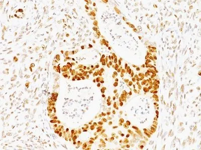
Recognizes a 53 kDa protein, which is identified as p53 suppressor gene product. It reacts with the mutant as well as the wild form of p53 protein. p53 is a tumor suppressor gene expressed in a wide variety of tissue types and is involved in regulating cell growth, replication, and apoptosis. It binds to MDM2, SV40 T antigen and human papilloma virus E6 protein. Positive nuclear staining with p53 antibody has been reported to be a negative prognostic factor in breast carcinoma, lung carcinoma, colorectal, and urothelial carcinoma. Anti-p53 positivity has also been used to differentiate uterine serous carcinoma from endometrioid carcinoma as well as to detect intratubular germ cell neoplasia. Mutations involving p53 are found in a wide variety of malignant tumors, including breast, ovarian, bladder, colon, lung, and melanoma.Primary antibodies are available purified, or with a selection of fluorescent CF® Dyes and other labels. CF® Dyes offer exceptional brightness and photostability. Note: Conjugates of blue fluorescent dyes like CF®405S and CF®405M are not recommended for detecting low abundance targets, because blue dyes have lower fluorescence and can give higher non-specific background than other dye colors.Synonyms:
Antigen NY-CO-13, BCC7, Cellular Tumor Antigen p53, LFS1, TP53, Transformation Related Protein 53 (TRP53), Tumor Protein p53, Tumor Suppressor p53CAS Number:
9007-83-4UNSPSC:
41116161UNSPSC Description:
Primary and secondary antibodies for multiple methodology immunostaining detection applicationGene Name:
TP53Gene ID:
7157NCBI Gene ID:
654481UniProt:
P04637Cellular Locus:
NucleusHost:
MouseSpecies Reactivity:
HumanImmunogen:
Recombinant human TP53 proteinTarget Antigen:
p53 Tumor Suppressor ProteinClonality:
MonoclonalIsotype:
IgG2b κClone:
TRP/817Conjugation:
CF640RSource:
AnimalApplications:
IHC, FFPE (verified) | WB (verified)Validated Applications:
IHC, FFPE, WBField of Research:
Apoptosis, Tumor suppressorsPositive Control:
MDA-MB-231 Cells. Breast or Colon carcinomaConcentration:
0.1 mg/mLBuffer:
PBS, 0.1% BSA, 0.05% azideMolecular Weight:
53 kDaAdditionnal Information:
Higher concentration may be required for direct detection using primary antibody conjugates than for indirect detection with secondary antibody|Immunofluorescence: 0.5-1 ug/mL|Immunohistology formalin-fixed 0.25-0.5 ug/mL|Staining of formalin-fixed tissues requires boiling tissue sections in 10 mM citrate buffer, pH 6.0, for 10-20 min followed by cooling at RT for 20 minutes|Flow Cytometry 0.5-1 ug/million cells/0.1 mL|Western blotting 0.5-1.0 ug/mL|Optimal dilution for a specific application should be determined by userShipping Conditions:
Room temperatureStorage Conditions:
4°C; Protect from light; Stable at room temperature or 37°C (98°F) for 7 days.Shelf Life:
2 years