Anti-p53(BP53-12), Biotin conjugate
CAT:
37-BNCB0013-500
Size:
500 µL
Price:
Ask
- Availability: 24/48H Stock Items & 2 to 6 Weeks non Stock Items.
- Dry Ice Shipment: No


Anti-p53(BP53-12), Biotin conjugate
Description:
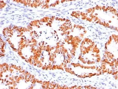
This MAb reacts with an N-terminal epitope (aa 16-25) of both wild type and mutated p53. Mutation and/or allelic loss of p53 is one of the causes of a variety of mesenchymal and epithelial tumors. If it occurs in the germ line, such tumors run in families. In most transformed and tumor cells the concentration of p53 is increased 51000 fold over the minute concentrations (1000 molecules cell) in normal cells, principally due to the increased half-life (4 h) compared to that of the wild-type (20 min). p53 Localizes in the nucleus, but is detectable at the plasma membrane during mitosis and when certain mutations modulate cytoplasmic/nuclear distribution. Mutations arise with an average frequency of 70% but incidence varies from zero in carcinoid lung tumors to 97% in primary melanomas. High concentrations of p53 protein are transiently expressed in human epidermis and superficial dermal fibroblasts following mild ultraviolet irradiation. Positive nuclear staining with p53 antibody has been reported to be a negative prognostic factor in breast carcinoma, lung carcinoma, colorectal, and urothelial carcinoma. Anti-p53 positivity has also been used to differentiate uterine serous carcinoma from endometrioid carcinoma as well as to detect intratubular germ cell neoplasia.Primary antibodies are available purified, or with a selection of fluorescent CF® Dyes and other labels. CF® Dyes offer exceptional brightness and photostability. Note: Conjugates of blue fluorescent dyes like CF®405S and CF®405M are not recommended for detecting low abundance targets, because blue dyes have lower fluorescence and can give higher non-specific background than other dye colors.Synonyms:
Antigen NY-CO-13, BCC7, Cellular Tumor Antigen p53, LFS1, TP53, Transformation Related Protein 53 (TRP53), Tumor Protein p53, Tumor Suppressor p53UNSPSC:
41116161UNSPSC Description:
Primary and secondary antibodies for multiple methodology immunostaining detection applicationGene Name:
TP53Gene ID:
7157NCBI Gene ID:
654481UniProt:
P04637Cellular Locus:
NucleusHost:
MouseSpecies Reactivity:
Chicken, Dog, Hamster, Human, MonkeyImmunogen:
Recombinant human wild-type p53 proteinTarget Antigen:
p53 Tumor Suppressor ProteinClonality:
MonoclonalIsotype:
IgG2aClone:
BP53-12Conjugation:
BiotinSource:
AnimalApplications:
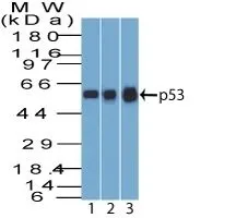
IHC, FFPE (verified) | WB (verified)Validated Applications:
IHC, FFPE, WBField of Research:
Apoptosis, Tumor suppressorsPositive Control:
MDA-MB-231 Cells. Breast or Colon carcinomaConcentration:
0.1 mg/mLBuffer:
PBS, 0.1% BSA, 0.05% azideMolecular Weight:
53 kDaAdditionnal Information:
Higher concentration may be required for direct detection using primary antibody conjugates than for indirect detection with secondary antibody|Immunofluorescence: 1-2 ug/mL|Does not react with mouse or rat, others not known|Immunohistology formalin-fixed 0.5-1 ug/mL|Staining of formalin-fixed tissues requires boiling tissue sections in 10 mM citrate buffer, pH 6.0, for 10-20 min followed by cooling at RT for 20 minutes|Flow Cytometry 0.5-1 ug/million cells/0.1 mL|Western blotting 0.5-1 ug/mL|Optimal dilution for a specific application should be determined by userShipping Conditions:
Room temperatureStorage Conditions:
4°C; Stable at room temperature or 37°C (98°F) for 7 days.Shelf Life:
2 yearsCAS Number:
9007-83-4
DATASHEET Document
View DocumentMSDS Document
View Document