
Anti-p27 / KIP1(DCS-72.F6), CF488A conjugate
CAT:
37-BNC880771-500
Size:
500 µL
Price:
Ask
- Availability: 24/48H Stock Items & 2 to 6 Weeks non Stock Items.
- Dry Ice Shipment: No


Anti-p27 / KIP1(DCS-72.F6), CF488A conjugate
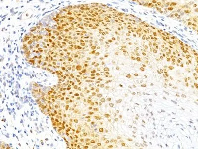
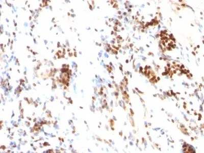
- Description: Recognizes a 27 kDa protein, identified as the p27Kip1, a cell cycle regulatory mitotic inhibitor. Its epitope spans between aa 83-204 of p27. It is highly specific and shows no cross-reaction with other related mitotic inhibitors. p27Kip1 functions as a negative regulator of G1 progression and has been proposed to function as a possible mediator of TGF-betanduced G1 arrest. p27Kip1 is a candidate tumor suppressor gene. This MAb co-precipitates cdk4 in complex p27Kip1 and is excellent for staining of formalin-fixed tissues.Primary antibodies are available purified, or with a selection of fluorescent CF® Dyes and other labels. CF® Dyes offer exceptional brightness and photostability. Note: Conjugates of blue fluorescent dyes like CF®405S and CF®405M are not recommended for detecting low abundance targets, because blue dyes have lower fluorescence and can give higher non-specific background than other dye colors.
- Synonyms: CDKN1B, CDKN4, Cyclin Dependent Kinase Inhibitor 1B, Cyclin-dependent kinase inhibitor p27 Kip1, KIP1, MEN1B, MEN4
- CAS Number: 9007-83-4
- UNSPSC: 41116161
- UNSPSC Description: Primary and secondary antibodies for multiple methodology immunostaining detection application
- Gene Name: CDKN1B
- Gene ID: 1027
- NCBI Gene ID: 238990
- UniProt: P46527
- Cellular Locus: Nucleus
- Host: Mouse
- Species Reactivity: Human, Monkey, Mouse, Rat
- Immunogen: Mouse recombinant p27 protein
- Target Antigen: KIP1 | p27
- Clonality: Monoclonal
- Isotype: IgG1 κ
- Clone: DCS-72.F6
- Conjugation: CF488A
- Source: Animal
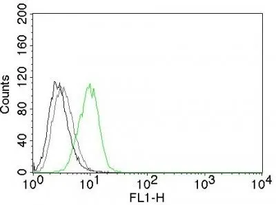
- Applications: Flow, intracellular (verified) | IHC, FFPE (verified)
- Validated Applications: FC, IHC, FFPE
- Field of Research: Cell cycle, Tumor suppressors
- Positive Control: ZR75, T47D, SK-BR-3, MDA-MB-231, HeLa or MCF7 cells. Tonsil, Breast, Cervical or Colon Carcinoma.
- Concentration: 0.1 mg/mL
- Buffer: PBS, 0.1% BSA, 0.05% azide
- Molecular Weight: 25-26 kDa
- Additionnal Information: Higher concentration may be required for direct detection using primary antibody conjugates than for indirect detection with secondary antibody|Immunofluorescence: 0.5-1 ug/mL|Immunohistology formalin-fixed 0.25-0.5 ug/mL|Staining of formalin-fixed tissues requires boiling tissue sections in 10 mM citrate buffer, pH 6.0, for 10-20 min followed by cooling at RT for 20 minutes|Flow Cytometry 0.5-1 ug/million cells/0.1 mL|Western blotting 0.5-1 ug/mL|Optimal dilution for a specific application should be determined by user
- Shipping Conditions: Room temperature
- Storage Conditions: 4°C; Protect from light; Stable at room temperature or 37°C (98°F) for 7 days.
- Shelf Life: 2 years