Anti-ODC-1 (Ornithine Decarboxylase-1)(ODC1/487), Biotin conjugate
CAT:
37-BNCB0487-500
Size:
500 µL
Price:
Ask
- Availability: 24/48H Stock Items & 2 to 6 Weeks non Stock Items.
- Dry Ice Shipment: No


Anti-ODC-1 (Ornithine Decarboxylase-1)(ODC1/487), Biotin conjugate
Description:
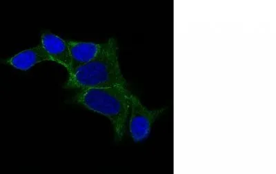
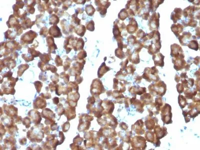
Recognizes a 53 kDa protein, identified as the Ornithine Decarboxylase (ODC-1). ODC is the initial and rate-limiting enzyme in the biosynthetic pathway of polyamines and is involved in the conversion of ornithine to putrescine. The biological activity of ODC-1 is rapidly induced in response to virtually all agents known to promote cell proliferation including hormones, drugs, growth factors, mitogens, and tumor promoters. Reportedly, ODC mRNA levels are elevated in lung carcinomas as well as in colon adenomas and carcinomas. ODC activity in colorectal carcinomas is greater than those in adenomas and normal mucosa.Primary antibodies are available purified, or with a selection of fluorescent CF® Dyes and other labels. CF® Dyes offer exceptional brightness and photostability. Note: Conjugates of blue fluorescent dyes like CF®405S and CF®405M are not recommended for detecting low abundance targets, because blue dyes have lower fluorescence and can give higher non-specific background than other dye colors.Synonyms:
Ornithine decarboxylase structural 1; RNODCUNSPSC:
41116161UNSPSC Description:
Primary and secondary antibodies for multiple methodology immunostaining detection applicationGene Name:
ODC1Gene ID:
4953NCBI Gene ID:
467701UniProt:
P11926Cellular Locus:
CytoplasmicHost:
MouseSpecies Reactivity:
Human, RatImmunogen:
Recombinant human ODC-1 proteinTarget Antigen:
Ornithine Decarboxylase 1Clonality:
MonoclonalIsotype:
IgG2a κClone:
ODC1/487Conjugation:
BiotinDisease:
TumorSource:
AnimalApplications:
Flow, intracellular (verified) | IF (verified)Validated Applications:
FC, IFField of Research:
Cancer, MetabolismPositive Control:
PC3 cells. Placenta, Prostate or Testicular carcinoma.Concentration:
0.1 mg/mLBuffer:
PBS, 0.1% BSA, 0.05% azideMolecular Weight:
53 kDaAdditionnal Information:
Higher concentration may be required for direct detection using primary antibody conjugates than for indirect detection with secondary antibody|Immunofluorescence: 0.5-1 ug/mL|Immunohistology (formalin)|Staining of formalin-fixed tissues requires boiling tissue sections in 10 mM citrate buffer, pH 6.0, for 10-20 min followed by cooling at RT for 20 minutes|Flow Cytometry 0.5-1 ug/million cells/0.1 mL|Western blotting 0.5-1 ug/mL|Optimal dilution for a specific application should be determined by userShipping Conditions:
Room temperatureStorage Conditions:
4°C; Stable at room temperature or 37°C (98°F) for 7 days.Shelf Life:
2 yearsCAS Number:
9007-83-4
DATASHEET Document
View DocumentMSDS Document
View Document