Anti-Myeloid-Associated Differentiation Marker(MYADM/971), CF568 conjugate
CAT:
37-BNC680971-100
Size:
100 µL
Price:
Ask
- Availability: 24/48H Stock Items & 2 to 6 Weeks non Stock Items.
- Dry Ice Shipment: No


Anti-Myeloid-Associated Differentiation Marker(MYADM/971), CF568 conjugate
Description:
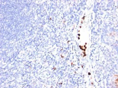
Recognizes a myeloid associated differentiation antigen in the cytoplasm of mature granulocytes. It shows no reactivity with any other cell type in human tissues. Markers of myeloid cells are useful in the identification of different levels of cellular differentiation. It reacts with early precursor and mature forms of human and monkey myeloid cells. This MAb is useful for the detection of myeloid leukemias and granulocytic sarcomas. It can be used as a marker of granulocytes in normal tissues or inflammatory processes.Primary antibodies are available purified, or with a selection of fluorescent CF® Dyes and other labels. CF® Dyes offer exceptional brightness and photostability. Note: Conjugates of blue fluorescent dyes like CF®405S and CF®405M are not recommended for detecting low abundance targets, because blue dyes have lower fluorescence and can give higher non-specific background than other dye colors.Synonyms:
MYADM; myeloid associated differentiation marker; Myeloid upregulated protein; Protein SB135CAS Number:
9007-83-4UNSPSC:
41116161UNSPSC Description:
Primary and secondary antibodies for multiple methodology immunostaining detection applicationGene ID:
91663NCBI Gene ID:
380906UniProt:
Q96S97Cellular Locus:
CytoplasmicHost:
MouseSpecies Reactivity:
Human, MacacaImmunogen:
Recombinant human MYADM proteinTarget Antigen:
Myeloid Associated Differentiation MarkerClonality:
MonoclonalIsotype:
IgG1Clone:
MYADM/971Conjugation:
CF568Source:
AnimalApplications:
IHC, FFPE (verified)Validated Applications:
IHC, FFPEField of Research:
ImmunologyPositive Control:
HL60 cells. Tonsil or lymph nodeConcentration:
0.1 mg/mLBuffer:
PBS, 0.1% BSA, 0.05% azideMolecular Weight:
UnknownAdditionnal Information:
Higher concentration may be required for direct detection using primary antibody conjugates than for indirect detection with secondary antibody|Immunofluorescence: 1-2 ug/mL|Immunohistology formalin-fixed 0.5-1 ug/mL|Staining of formalin-fixed tissues requires boiling tissue sections in 10 mM citrate buffer, pH 6.0, for 10-20 min followed by cooling at RT for 20 minutes|Flow Cytometry 0.5-1 ug/million cells/0.1 mL|Optimal dilution for a specific application should be determined by userShipping Conditions:
Room temperatureStorage Conditions:
4°C; Protect from light; Stable at room temperature or 37°C (98°F) for 7 days.Shelf Life:
2 years
DATASHEET Document
View DocumentMSDS Document
View Document