Anti-MART-1 / Melan-A(M2-7C10 + M2-9E3 + A103), CF640R conjugate
CAT:
37-BNC400700-100
Size:
100 µL
Price:
Ask
- Availability: 24/48H Stock Items & 2 to 6 Weeks non Stock Items.
- Dry Ice Shipment: No


Anti-MART-1 / Melan-A(M2-7C10 + M2-9E3 + A103), CF640R conjugate
Description:
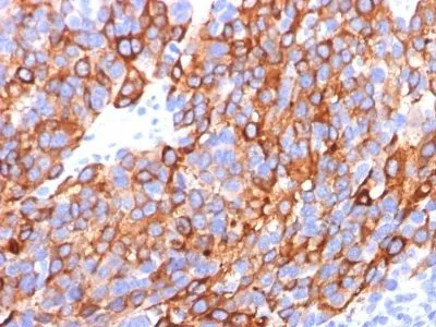
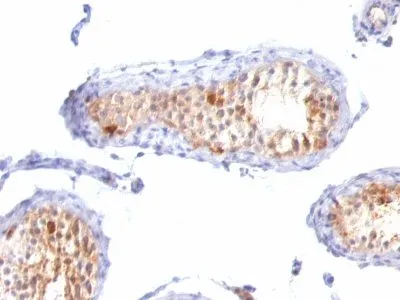
This antibody recognizes a protein doublet of 20-22 kDa, identified as MART-1 (Melanoma Antigen Recognized by T cells 1) or Melan-A. MART-1 is a newly identified melanocyte differentiation antigen recognized by autologous cytotoxic T lymphocytes. Seven other melanoma associated antigens recognized by autologous cytotoxic T cells include MAGE-1, MAGE-3, tyrosinase, gp100, gp75, BAGE-1, and GAGE-1. Subcellular fractionation shows that MART-1 is present in melanosomes and endoplasmic reticulum. This MAb cocktail labels melanomas and other tumors showing melanocytic differentiation. It is also a useful positive-marker for angiomyolipomas. It does not stain tumor cells of epithelial, lymphoid, glial, or mesenchymal origin.Primary antibodies are available purified, or with a selection of fluorescent CF® Dyes and other labels. CF® Dyes offer exceptional brightness and photostability. Note: Conjugates of blue fluorescent dyes like CF®405S and CF®405M are not recommended for detecting low abundance targets, because blue dyes have lower fluorescence and can give higher non-specific background than other dye colors.Synonyms:
Antigen LB39-AA, Antigen SK29-AA, Melanoma antigen recognized by T-cells 1, MLAN-A, MLANAUNSPSC:
41116161UNSPSC Description:
Primary and secondary antibodies for multiple methodology immunostaining detection applicationGene Name:
MLANAGene ID:
2315NCBI Gene ID:
154069UniProt:
Q16655Cellular Locus:
Golgi apparatusHost:
MouseSpecies Reactivity:
Dog, Human, Mouse, RatImmunogen:
Recombinant hMART-1 protein (A103; M2-7C10; M2-9E3)Target Antigen:
MART-1 | Melan-AClonality:
MonoclonalIsotype:
IgGClone:
M2-7C10 M2-9E3 A103Conjugation:
CF640RDisease:
TumorSource:
AnimalApplications:
IHC, FFPE (verified)Validated Applications:
IHC, FFPEField of Research:
CancerPositive Control:
SK-MEL-13 and SK-MEL-19 Melanoma cell lines; MelanomasConcentration:
0.1 mg/mLBuffer:
PBS, 0.1% BSA, 0.05% azideMolecular Weight:
20-22 kDa (doublet)Additionnal Information:
Higher concentration may be required for direct detection using primary antibody conjugates than for indirect detection with secondary antibody|Immunofluorescence: 0.5-1 ug/mL|Immunohistology formalin-fixed 0.5-1 ug/mL|Staining of formalin-fixed tissues is enhanced by boiling tissue sections in 10 mM citrate buffer, pH 6.0, for 10-20 min followed by cooling at RT for 20 minutes|Flow Cytometry 0.5-1 ug/million cells/0.1 mL|Western blotting 0.5-1.0 ug/mL|Optimal dilution for a specific application should be determined by userShipping Conditions:
Room temperatureStorage Conditions:
4°C; Protect from light; Stable at room temperature or 37°C (98°F) for 7 days.Shelf Life:
2 yearsCAS Number:
9007-83-4
DATASHEET Document
View DocumentMSDS Document
View Document