
Anti-MAGE A1(MZ2E/838)
CAT:
37-BNUB0838-500
Size:
500 µL
Price:
Ask
- Availability: 24/48H Stock Items & 2 to 6 Weeks non Stock Items.
- Dry Ice Shipment: No


Anti-MAGE A1(MZ2E/838)
- Description: Recognizes a protein of 42-46 kDa, identified as MAGE-1. This MAb does not cross-react with other members of MAGE-family. Human malignant neoplasms carry rejection antigens that are recognized by the patients' autologous, tumor directed and specific, cytolytic, CD8 T lymphocyte clones (CTL). The MAGE family of genes codes an important group of antigens. It was identified that melanomas and primary glial brain tumors express common melanoma associated antigens (MAAs). Because MAGE-1 is expressed on a significant proportion of human neoplasms of various histological types (melanoma, brain tumors of glial origin, neuroblastoma, non-small cell lung cancer, breast, gastric, colorectal, ovarian, renal cell carcinomas) and not on normal tissues, the encoded antigen may serve as a marker of early detection and target for cancer immunotherapy.Primary antibodies are available purified, or with a selection of fluorescent CF® Dyes and other labels. CF® Dyes offer exceptional brightness and photostability. Note: Conjugates of blue fluorescent dyes like CF®405S and CF®405M are not recommended for detecting low abundance targets, because blue dyes have lower fluorescence and can give higher non-specific background than other dye colors.
- Synonyms: MZ2 E, cancer/testis antigen 1.1, CT1.1, MAGE1A, MAGEA1, Melanoma antigen family A 1, Melanoma associated antigen 1, Melanoma associated antigen MZ2 E
- CAS Number: 9007-83-4
- UNSPSC: 41116161
- UNSPSC Description: Primary and secondary antibodies for multiple methodology immunostaining detection application
- Gene Name: MAGEA1
- Gene ID: 4100
- NCBI Gene ID: 72879
- UniProt: P43355
- Cellular Locus: Cytoplasmic
- Host: Mouse
- Species Reactivity: Human
- Immunogen: Recombinant human MAGEA1 protein
- Target Antigen: MAGE-A1
- Clonality: Monoclonal
- Isotype: IgG1 κ
- Clone: MZ2E/838
- Conjugation: Purified, with BSA
- Disease: Tumor
- Source: Animal
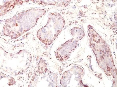
- Applications: IHC, FFPE (verified)
- Validated Applications: IHC, FFPE
- Field of Research: Cancer
- Positive Control: Melanoma cell lines. Melanomas, gliomas, neuroblastoma, non-small cell lung cancer, breast, gastric, colorectal, ovarian, and renal cell carcinomas.
- Concentration: 0.2 mg/mL
- Buffer: PBS, 0.05% BSA, 0.05% azide
- Molecular Weight: 42-46 kDa
- Additionnal Information: Higher concentration may be required for direct detection using primary antibody conjugates than for indirect detection with secondary antibody|Immunofluorescence: 1-2 ug/mL|Immunohistology formalin-fixed 0.5-1 ug/mL|Staining of formalin-fixed tissues requires boiling tissue sections in 10 mM citrate buffer, pH 6.0, for 10-20 min followed by cooling at RT for 20 minutes|Flow Cytometry 0.5-1 ug/million cells/0.1 mL|Optimal dilution for a specific application should be determined by user
- Shipping Conditions: Room temperature
- Storage Conditions: 4°C; Stable at room temperature or 37°C (98°F) for 7 days.
- Shelf Life: 2 years