Anti-HSP60(HSPD1/780)
CAT:
37-BNUB0780-500
Size:
500 µL
Price:
Ask
- Availability: 24/48H Stock Items & 2 to 6 Weeks non Stock Items.
- Dry Ice Shipment: No


Anti-HSP60(HSPD1/780)
Description:
Recognizes a 60 kDa protein, identified as the heat shock protein 60 (hsp60). A wide variety of environmental and pathophysiological stressful conditions trigger the synthesis of a family of proteins known as heat shock proteins (HSPs), more appropriately called as stress response proteins (SRPs). Hsp60 is a potential antigen in a number of autoimmune diseases. In human arthritis and in experimentally induced arthritis in animals, disease development coincides with the development of immune reactivity directed against not only bacterial hsp60, but also against its mammalian homolog.Primary antibodies are available purified, or with a selection of fluorescent CF® Dyes and other labels. CF® Dyes offer exceptional brightness and photostability. Note: Conjugates of blue fluorescent dyes like CF®405S and CF®405M are not recommended for detecting low abundance targets, because blue dyes have lower fluorescence and can give higher non-specific background than other dye colors.Synonyms:
60kDa chaperonin; 60kDa heat shock protein mitochondrial; Chaperonin; 60-KD (CPN60); GROEL; HLD4; HSP65; HSPD1; HuCHA60; Mitochondrial matrix protein P1; P60 lymphocyte protein; Short heat shock protein 60 Hsp60s1; Spastic paraplegia 13 (SPG13)UNSPSC:
41116161UNSPSC Description:
Primary and secondary antibodies for multiple methodology immunostaining detection applicationGene Name:
HSPD1Gene ID:
3329NCBI Gene ID:
595053UniProt:
P10809Cellular Locus:
MitochondriaHost:
MouseSpecies Reactivity:
Chicken, Cow, Dog, Hamster, Human, Monkey, Mouse, Pig, Rabbit, Rat, SheepImmunogen:
Recombinant human HSPD1 proteinTarget Antigen:
HSP60Clonality:
MonoclonalIsotype:
IgG1 κClone:
HSPD1/780Conjugation:
Purified, with BSASource:
AnimalApplications:
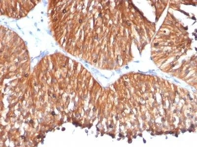
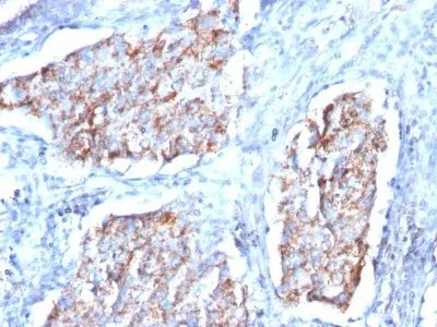
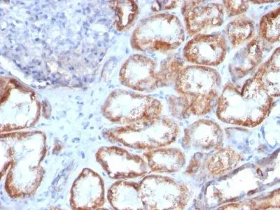
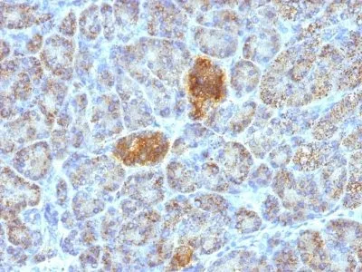
IF (verified) | IHC, FFPE (verified) | WB (verified)Validated Applications:
IF, IHC, FFPE, WBField of Research:
Autoimmunity, Heat shock proteins, Immunology, InflammationPositive Control:
HeLa or HepG2 cells. Synovial biopsies from patients with juvenile chronic arthritis. Synovial lining layer is strongly positive for hsp60. Breast carcinoma.Concentration:
0.2 mg/mLBuffer:
PBS, 0.05% BSA, 0.05% azideMolecular Weight:
60 kDaAdditionnal Information:
Higher concentration may be required for direct detection using primary antibody conjugates than for indirect detection with secondary antibody|Immunofluorescence: 0.5-1 ug/mL|Immunohistology formalin-fixed 0.5-1 ug/mL|Staining of formalin-fixed tissues is enhanced by boiling tissue sections in 10 mM citrate buffer, pH 6.0, for 10-20 min followed by cooling at RT for 20 minutes|Flow Cytometry 0.5-1 ug/million cells/0.1 mL|Western blotting 0.25-0.5 ug/mL|Optimal dilution for a specific application should be determined by userShipping Conditions:
Room temperatureStorage Conditions:
4°C; Stable at room temperature or 37°C (98°F) for 7 days.Shelf Life:
2 yearsCAS Number:
9007-83-4
DATASHEET Document
View DocumentMSDS Document
View Document