
Anti-Hepatocyte Specific(OCH1E5)
- Availability: 24/48H Stock Items & 2 to 6 Weeks non Stock Items.
- Dry Ice Shipment: No


Anti-Hepatocyte Specific(OCH1E5)
Description:
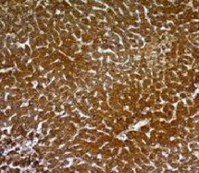
Hepatocyte Specific Antigen, also called Hepatocyte Paraffin 1 or HepPar1, localizes to the mitochondria of hepatocytes. It is a sensitive marker for distinguishing hepatocellular carcinomas (HCC) from other metastatic carcinomas as well as cholangio-carcinomas. HCC's occur primarily in the stomach, but they are also found in many other organs. The Hepatocyte Specific Antigen may also be a useful marker for intestinal metaplasia. Reportedly, strong expression of the Hepatocyte Specific Antigen correlates with smaller tumor size and longer patient survival. Occasionally, Hepatocyte Specific Antigen is also found in gastric carcinomas as well as in a few other non-hepatic tumors._x000D__x000D_Primary antibodies are available purified, or with a selection of fluorescent CF® Dyes and other labels. CF® Dyes offer exceptional brightness and photostability. Note: Conjugates of blue fluorescent dyes like CF®405S and CF®405M are not recommended for detecting low abundance targets, because blue dyes have lower fluorescence and can give higher non-specific background than other dye colors.Synonyms:
Not KnownUNSPSC:
41116161UNSPSC Description:
Primary and secondary antibodies for multiple methodology immunostaining detection applicationGene ID:
Not KnownNCBI Gene ID:
Not KnownUniProt:
Not KnownCellular Locus:
CytoplasmicHost:
MouseSpecies Reactivity:
Dog, HumanImmunogen:
Extract of a formalin-fixed, rejected-allograft of a human liverTarget Antigen:
Hepatocyte Specific AntigenClonality:
MonoclonalIsotype:
IgG1Clone:
OCH1E5Conjugation:
Purified, with BSADisease:
TumorSource:
AnimalApplications:
IHC, FFPE (verified)Validated Applications:
IHC, FFPEField of Research:
Cancer, Signal transductionPositive Control:
Liver or Hepatocellular Carcinoma (HCC)Concentration:
0.2 mg/mLBuffer:
PBS, 0.05% BSA, 0.05% azideMolecular Weight:
Not KnownAdditionnal Information:
Immunohistology formalin-fixed 1-2 ug/mL|Staining of formalin-fixed tissues requires boiling tissue sections in 10 mM Tris with 1 mM EDTA, pH 9.0, for 10-20 min followed by cooling at RT for 20 minutes|Immunofluorescence 0.5-1.0 ug/mL|Optimal dilution for a specific application should be determined by userShipping Conditions:
Room temperatureStorage Conditions:
4°C; Stable at room temperature or 37°C (98°F) for 7 days.Shelf Life:
2 yearsCAS Number:
9007-83-4
DATASHEET Document
View DocumentMSDS Document
View Document