
Anti-DOG-1(DOG1.1), Biotin conjugate
CAT:
37-BNCB0725-100
Size:
100 µL
Price:
Ask
- Availability: 24/48H Stock Items & 2 to 6 Weeks non Stock Items.
- Dry Ice Shipment: No


Anti-DOG-1(DOG1.1), Biotin conjugate
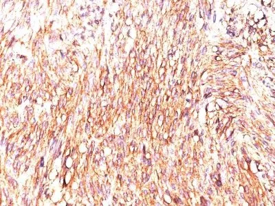
- Description: Expression of DOG-1 protein is elevated in the gastrointestinal stromal tumors (GISTs), c-kit signaling-driven mesenchymal tumors of the GI tract. DOG-1 is rarely expressed in other soft tissue tumors, which, due to appearance, may be difficult to diagnose. Immunoreactivity for DOG-1 has been reported in 97.8 percent of scorable GISTs, including all c-kit negative GISTs. Overexpression of DOG-1 has been suggested to aid in the identification of GISTs, including Platelet-Derived Growth Factor Receptor Alpha mutants that fail to express c-kit antigen. The overall sensitivity of DOG1 and c-kit in GISTs is nearly identical: 94.4% vs. 94.7%.Primary antibodies are available purified, or with a selection of fluorescent CF® Dyes and other labels. CF® Dyes offer exceptional brightness and photostability. Note: Conjugates of blue fluorescent dyes like CF®405S and CF®405M are not recommended for detecting low abundance targets, because blue dyes have lower fluorescence and can give higher non-specific background than other dye colors.
- Synonyms: Anoctamin 1; Calcium Activated Chloride Channel; Discovered On Gastrointestinal Stromal Tumors Protein 1; TAOS2; ORAOV2; TMEM16A
- CAS Number: 9007-83-4
- UNSPSC: 41116161
- UNSPSC Description: Primary and secondary antibodies for multiple methodology immunostaining detection application
- Gene Name: TMEM16A
- Gene ID: 55107
- NCBI Gene ID: 503074
- UniProt: Q5XXA6
- Cellular Locus: Plasma membrane|Nucleus
- Host: Mouse
- Species Reactivity: Human
- Immunogen: A synthetic peptide from human DOG-1 protein (MSDFVDWVIPDIPKDISQQIHKEKVLMVELFMREEQDKQQL-LETCMEKERQKDEPPCNHHNTKACPDSLGSPAPSHAYHGGVL), conjugated to a carrier protein.
- Target Antigen: DOG-1 | TMEM16A
- Clonality: Monoclonal
- Isotype: IgG1 κ
- Clone: DOG1.1
- Conjugation: Biotin
- Disease: Tumor
- Source: Animal
- Applications: IHC, FFPE (verified)
- Validated Applications: IHC, FFPE
- Field of Research: Cancer
- Positive Control: Gastrointestinal Stromal Tumor (GIST) or testicular germ cell tumor. Melanocytes in the basal layer of the epidermis and mast cells in the dermis of normal skin.
- Concentration: 0.1 mg/mL
- Buffer: PBS, 0.1% BSA, 0.05% azide
- Molecular Weight: ~114 kDa
- Additionnal Information: Higher concentration may be required for direct detection using primary antibody conjugates than for indirect detection with secondary antibody|Immunofluorescence: 0.5-1 ug/mL|Immunohistology formalin-fixed 0.25-0.5 ug/mL|Staining of formalin-fixed tissues requires boiling tissue sections in 10 mM citrate buffer, pH 6.0, for 10-20 min followed by cooling at RT for 20 minutes|Flow Cytometry 0.5-1 ug/million cells/0.1 mL|Optimal dilution for a specific application should be determined by user
- Shipping Conditions: Room temperature
- Storage Conditions: 4°C; Stable at room temperature or 37°C (98°F) for 7 days.
- Shelf Life: 2 years