
Anti-Cytokeratin 7(K72.7)
- Availability: 24/48H Stock Items & 2 to 6 Weeks non Stock Items.
- Dry Ice Shipment: No


Anti-Cytokeratin 7(K72.7)
Description:
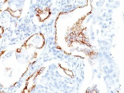
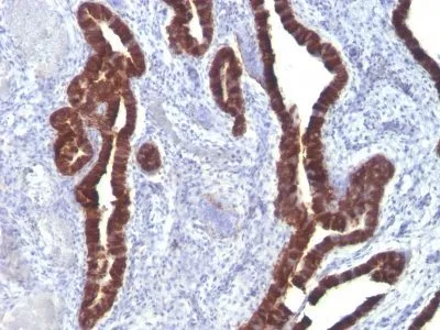
This antibody recognizes an intermediate filament protein (IFP) of 55 kDa, which is identified as cytokeratin 7. This MAb is highly specific to cytokeratin 7 and shows no cross-reaction with other IFPs. Cytokeratin 7 is a basic cytokeratin, which is found in most glandular and transitional epithelia but not in the stratified squamous epithelia. Keratin 7 is expressed in the epithelial cells of ovary, lung, and breast but not of colon, prostate, or gastrointestinal tract. This MAb is highly useful in distinguishing ovarian carcinomas (keratin 7 ) from colon carcinomas (keratin 7-).Primary antibodies are available purified, or with a selection of fluorescent CF® Dyes and other labels. CF® Dyes offer exceptional brightness and photostability. Note: Conjugates of blue fluorescent dyes like CF®405S and CF®405M are not recommended for detecting low abundance targets, because blue dyes have lower fluorescence and can give higher non-specific background than other dye colors.Synonyms:
CK-7, K2C7, Keratin 55K Type II Cytoskeletal, Keratin Simple Epithelial Type 1 K7, Keratin Type II Cytoskeletal 7, Krt2-7, KRT7, Sarcolectin, SCL, Type II Mesothelial Keratin K7, Type-II Keratin Kb7UNSPSC:
41116161UNSPSC Description:
Primary and secondary antibodies for multiple methodology immunostaining detection applicationGene Name:
KRT7Gene ID:
3855NCBI Gene ID:
411501UniProt:
P08729Cellular Locus:
CytoskeletonHost:
MouseSpecies Reactivity:
HumanImmunogen:
Semi-purified cytokeratin preparation.Target Antigen:
Cytokeratin 7Clonality:
MonoclonalIsotype:
IgG1 κClone:
K72.7Conjugation:
Purified, BSA-freeDisease:
TumorSource:
AnimalApplications:
Flow, intracellular (verified) | IF (verified) | IHC, FFPE (verified) | WB (verified)Validated Applications:
FC, IF, IHC, FFPE, WBField of Research:
Cancer, CytoskeletonPositive Control:
HeLa cells. Carcinoma of ovary, lung, cervix, or breastConcentration:
1 mg/mLBuffer:
PBS, no BSA, no azideMolecular Weight:
55 kDaAdditionnal Information:
Higher concentration may be required for direct detection using primary antibody conjugates than for indirect detection with secondary antibody|Immunofluorescence: 0.5-1 ug/mL|Immunohistology (formalin)|Staining of formalin-fixed tissues requires boiling tissue sections in 10 mM citrate buffer, pH 6.0, for 10-20 min followed by cooling at RT for 20 minutes|Flow Cytometry 0.5-1 ug/million cells/0.1 mL|Optimal dilution for a specific application should be determined by userShipping Conditions:
Room temperatureStorage Conditions:
-35°C to -5°C ; Stable at room temperature or 37°C (98°F) for 7 days.Shelf Life:
2 yearsCAS Number:
9007-83-4
DATASHEET Document
View DocumentMSDS Document
View Document