
Anti-CD79a(HM47/A9), CF640R conjugate
CAT:
37-BNC400477-100
Size:
100 µL
Price:
Ask
- Availability: 24/48H Stock Items & 2 to 6 Weeks non Stock Items.
- Dry Ice Shipment: No


Anti-CD79a(HM47/A9), CF640R conjugate
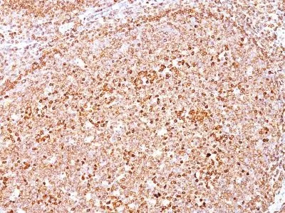
- Description: CD79 is a disulphide-linked heterodimer, consisting of mb-1 (or CD79a) and B29 (or CD79b) polypeptides, is non-covalently associated with membrane-bound immunoglobulins on B cells. This complex of mb-1 and B29 polypeptides and immunoglobulin constitute the B cell Ag receptor. CD79a first appears at pre B cell stage, early in maturation, and persists until the plasma cell stage where it is found as an intracellular component. CD79a is found in the majority of acute leukemias of precursor B cell type, in B cell lines, B cell lymphomas, and in some myelomas. It is not present in myeloid or T cell lines. Anti-CD79a is generally used to complement anti-CD20 especially for mature B-cell lymphomas after treatment with Rituximab (anti-CD20). This antibody will stain many of the same lymphomas as anti-CD20, but also is more likely to stain B-lymphoblastic lymphoma/leukemia than is anti-CD20. Anti-CD79a also stains more cases of plasma cell myeloma and occasionally some types of endothelial cells as well.Primary antibodies are available purified, or with a selection of fluorescent CF® Dyes and other labels. CF® Dyes offer exceptional brightness and photostability. Note: Conjugates of blue fluorescent dyes like CF®405S and CF®405M are not recommended for detecting low abundance targets, because blue dyes have lower fluorescence and can give higher non-specific background than other dye colors.
- Synonyms: B lymphocyte-specific MB1 protein|B-cell antigen receptor complex-associated protein alpha chain|CD79a molecule immunoglobulin associated alpha|Ig-alpha|IGA|IgM-alpha|Immunoglobulin-associated alpha|Ly54|MB-1 membrane glycoprotein|Membrane-bound immunoglobulin-associated protein|Surface IgM-associated protein
- CAS Number: 9007-83-4
- UNSPSC: 41116161
- UNSPSC Description: Primary and secondary antibodies for multiple methodology immunostaining detection application
- Gene Name: CD79A
- Gene ID: 973
- NCBI Gene ID: 631567
- UniProt: P11912
- Cellular Locus: Plasma membrane
- Host: Mouse
- Species Reactivity: Cow, Human, Monkey, Mouse, Pig, Rat
- Immunogen: A synthetic peptide corresponding to aa 202-216 (GTYQDVGSLNIADVQ) of human CD79a protein.
- Target Antigen: CD79a
- Clonality: Monoclonal
- Isotype: IgG1 κ
- Clone: HM47/A9
- Conjugation: CF640R
- Disease: Tumor
- Source: Animal
- Applications: Flow (verified) | IF (verified) | IHC, FFPE (verified) | WB (verified)
- Validated Applications: FC, IF, IHC, FFPE, WB
- Field of Research: Immunology
- Positive Control: Daudi or Ramos cells. Germinal center B-cells in a lymph node or tonsil.
- Concentration: 0.1 mg/mL
- Buffer: PBS, 0.1% BSA, 0.05% azide
- Molecular Weight: 44 kDa
- Additionnal Information: Higher concentration may be required for direct detection using primary antibody conjugates than for indirect detection with secondary antibody|Immunofluorescence: 0.5-1 ug/mL|Immunohistology formalin-fixed 0.25-0.5 ug/mL|Staining of formalin-fixed tissues requires boiling tissue sections in 10 mM citrate buffer, pH 6.0, for 10-20 min followed by cooling at RT for 20 minutes|Flow Cytometry 0.5-1 ug/million cells/0.1 mL|Optimal dilution for a specific application should be determined by user
- Shipping Conditions: Room temperature
- Storage Conditions: 4°C; Protect from light; Stable at room temperature or 37°C (98°F) for 7 days.
- Shelf Life: 2 years