
Anti-CD68(CD68/G2)
CAT:
37-BNUM0919-50
Size:
50 µL
Price:
Ask
- Availability: 24/48H Stock Items & 2 to 6 Weeks non Stock Items.
- Dry Ice Shipment: No
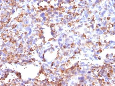


Anti-CD68(CD68/G2)
- Description: This antibody recognizes a glycoprotein of 110 kDa, which is identified as CD68. It is important for identifying macrophages in tissue sections. It stains macrophages in a wide variety of human tissues, including Kupffer cells and macrophages in the red pulp of the spleen, in lamina propria of the gut, in lung alveoli, and in bone marrow. It reacts with myeloid precursors and peripheral blood granulocytes. It also reacts with plasmacytoid T cells, which are supposed to be of monocyte/macrophage origin. It shows strong granular cytoplasmic staining of chronic and acute myeloid leukemia and also reacts with rare cases of true histiocytic neoplasia. Lymphomas are negative or show few granules.Primary antibodies are available purified, or with a selection of fluorescent CF® Dyes and other labels. CF® Dyes offer exceptional brightness and photostability. Note: Conjugates of blue fluorescent dyes like CF®405S and CF®405M are not recommended for detecting low abundance targets, because blue dyes have lower fluorescence and can give higher non-specific background than other dye colors.
- Synonyms: GP110, LAMP4, Microsialin, Macrosialin, SCARD1, Scavenger Receptor Class D Member-1
- CAS Number: 9007-83-4
- UNSPSC: 41116161
- UNSPSC Description: Primary and secondary antibodies for multiple methodology immunostaining detection application
- Gene Name: CD68
- Gene ID: 968
- NCBI Gene ID: 647419
- UniProt: P34810
- Cellular Locus: Plasma membrane
- Host: Mouse
- Species Reactivity: Human
- Immunogen: Recombinant human CD68 protein
- Target Antigen: CD68
- Clonality: Monoclonal
- Isotype: IgG1 κ
- Clone: CD68/G2
- Conjugation: Purified, BSA-free
- Disease: Tumor
- Source: Animal
- Applications: IHC, FFPE (verified)
- Validated Applications: IHC, FFPE
- Field of Research: Immunology
- Positive Control: Tonsil, lymph node, or spleen
- Concentration: 1 mg/mL
- Buffer: PBS, no BSA, no azide
- Molecular Weight: ~110 kDa
- Additionnal Information: Higher concentration may be required for direct detection using primary antibody conjugates than for indirect detection with secondary antibody|Immunofluorescence: 0.5-1 ug/mL|Immunohistology formalin-fixed 0.5-1 ug/mL|Staining of formalin-fixed tissues is enhanced by boiling tissue sections in 10 mM citrate buffer, pH 6.0, for 10-20 min followed by cooling at RT for 20 minutes|Flow Cytometry 0.5-1 ug/million cells/0.1 mL|Optimal dilution for a specific application should be determined by user
- Shipping Conditions: Room temperature
- Storage Conditions: -35°C to -5°C ; Stable at room temperature or 37°C (98°F) for 7 days.
- Shelf Life: 2 years