Anti-CD6(3F7B5), CF640R conjugate
CAT:
37-BNC400428-100
Size:
100 µL
Price:
Ask
- Availability: 24/48H Stock Items & 2 to 6 Weeks non Stock Items.
- Dry Ice Shipment: No


Anti-CD6(3F7B5), CF640R conjugate
Description:
CD6 is a type I transmembrane glycoprotein that contains a 24-amino acid signal sequence, three extracellular scavenger receptor cysteine-rich (SRCR) domains, a membrane-spanning domain and a 44-amino acid cytoplasmic domain. The CD6 glycoprotein is tyrosine phosphorylated during TCR-mediated T cell activation. CD6 shows significant homology to CD5. CD6 is present on mature thymocytes, peripheral T cells and a subset of B cells. Antibodies to CD6 are used to deplete T cells from bone marrow transplants to prevent graft versus host disease.Primary antibodies are available purified, or with a selection of fluorescent CF® Dyes and other labels. CF® Dyes offer exceptional brightness and photostability. Note: Conjugates of blue fluorescent dyes like CF®405S and CF®405M are not recommended for detecting low abundance targets, because blue dyes have lower fluorescence and can give higher non-specific background than other dye colors.Synonyms:
T12; TP120CAS Number:
9007-83-4UNSPSC:
41116161UNSPSC Description:
Primary and secondary antibodies for multiple methodology immunostaining detection applicationGene Name:
CD6Gene ID:
923NCBI Gene ID:
744366UniProt:
P30203Cellular Locus:
Plasma membraneHost:
MouseSpecies Reactivity:
HumanImmunogen:
Human rheumatoid synovial T cell line ST-1Target Antigen:
CD6Clonality:
MonoclonalIsotype:
IgG1Clone:
3F7B5Conjugation:
CF640RSource:
AnimalApplications:
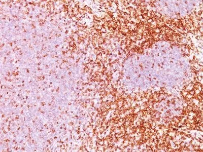
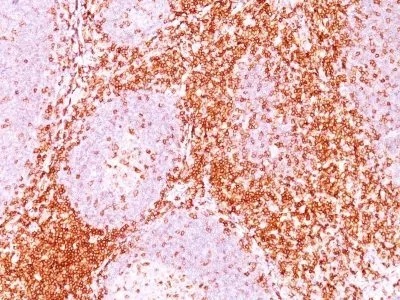
Flow (verified) | IF (verified) | IHC, FFPE (verified)Validated Applications:
FC, IF, IHC, FFPEField of Research:
ImmunologyPositive Control:
CCRF-CEM, Jurkat cells, TonsilConcentration:
0.1 mg/mLBuffer:
PBS, 0.1% BSA, 0.05% azideMolecular Weight:
90-130 kDaAdditionnal Information:
Higher concentration may be required for direct detection using primary antibody conjugates than for indirect detection with secondary antibody|Immunofluorescence: 0.5-1 ug/mL|Immunohistology formalin-fixed 0.5-1 ug/mL|Staining of formalin-fixed tissues requires boiling tissue sections in 10 mM citrate buffer, pH 6.0, for 10-20 min followed by cooling at RT for 20 minutes|Flow Cytometry 0.5-1 ug/million cells/0.1 mL|Optimal dilution for a specific application should be determined by userShipping Conditions:
Room temperatureStorage Conditions:
4°C; Protect from light; Stable at room temperature or 37°C (98°F) for 7 days.Shelf Life:
2 years