Anti-CD57 / B3GAT1(NK-1)
CAT:
37-BNUM0016-50
Size:
50 µL
Price:
Ask
- Availability: 24/48H Stock Items & 2 to 6 Weeks non Stock Items.
- Dry Ice Shipment: No


Anti-CD57 / B3GAT1(NK-1)
Description:
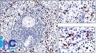
Anti-CD57 marks a subset of lymphocytes known as natural killer (NK) cells. Follicular center cell lymphomas often contain many NK cells within the neoplastic follicles. Anti-CD57 also stains neuroendocrine cells and their derived tumors, including carcinoid tumor and medulloblastoma. Anti-CD57 can also be useful in separating type B3 thymoma from thymic carcinoma when combined with a panel that includes antibodies against GLUT1, CD5, and CEA._x000D_ _x000D_ This antibody is available purified with BSA/azide at 200 ug/mL, or BSA/azide-free at 1 mg/mL.Synonyms:
3-Glucuronyltransferase 1; B3GAT1; Galactosylgalactosylxylosylprotein 3-beta-Glucuronosyltransferase 1; GLCATP; GlcUAT-P; Glucuronosyltransferase P; UDP GlcUA Glycoprotein beta 1, 3 GlucuronyltransferaseUNSPSC:
41116161UNSPSC Description:
Primary and secondary antibodies for multiple methodology immunostaining detection applicationGene Name:
B3GAT1Gene ID:
27087NCBI Gene ID:
381050UniProt:
Q9P2W7Cellular Locus:
Plasma membraneHost:
MouseSpecies Reactivity:
HumanImmunogen:
Human peripheral blood mononuclear cellsTarget Antigen:
B3GAT1 | CD57Clonality:
MonoclonalIsotype:
IgM κClone:
NK-1Conjugation:
Purified, BSA-freeSource:
AnimalApplications:
Flow, surface (published) | IF (published) | IHC, FFPE (verified) | WB (published)Validated Applications:
FC, IF, IHC, FFPE, WBField of Research:
Cancer, ImmunologyPositive Control:
Lymph node or TonsilConcentration:
1 mg/mLBuffer:
PBS, no BSA, no azideMolecular Weight:
~110 kDa (Glycoprotein)Additionnal Information:
Higher concentration may be required for direct detection using primary antibody conjugates than for indirect detection with secondary antibody|Immunofluorescence: 0.5-1 ug/mL|Does not react with rat; others not known|Immunohistology formalin-fixed 2-4 ug/mL|Staining of formalin-fixed tissues requires boiling tissue sections in 10 mM Tris with 1 mM EDTA, pH 9.0, for 10-20 min followed by cooling at RT for 20 minutes|Optimal dilution for a specific application should be determined by userReferences & Citations:
Note: References for this clone sold by other suppliers may be listed for expected applications. Asian J Androl (2010) 12(4): 548-555. (WB blot; Flow) Cancer Immunol Immunother (2011) 60:1683-1695. (Flow, surface) Cancer Sci (2016) 107: 846-852. (IHC, FFPE) Cell Stem Cell (2017) 20(6): 874-890.e7. (IF; Flow, surface)Shipping Conditions:
Room temperatureStorage Conditions:
-35°C to -5°C ; Stable at room temperature or 37°C (98°F) for 7 days.Shelf Life:
2 yearsCAS Number:
9007-83-4
DATASHEET Document
View DocumentMSDS Document
View Document