Anti-GLG1 (Golgi Complex)(GLG1/970), CF740 conjugate
CAT:
37-BNC740970-500
Size:
500 µL
Price:
Ask
- Availability: 24/48H Stock Items & 2 to 6 Weeks non Stock Items.
- Dry Ice Shipment: No


Anti-GLG1 (Golgi Complex)(GLG1/970), CF740 conjugate
Description:
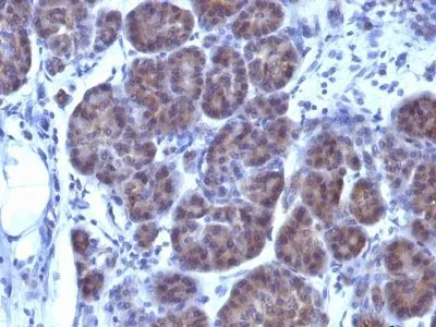
This MAb recognizes a protein of 134 kDa, which binds fibroblast growth factor and E-selectin (cell-adhesion lectin on endothelial cells mediating the binding of neutrophils). Fucosylation is essential for binding to E-selectin. It contains sialic acid residues and 16 Cys-rich GLG1 repeats. This MAb can be used to stain the Golgi complex in cell or tissue preparations and can be used as a Golgi marker in subcellular fractions. It produces a diffuse staining pattern of the Golgi zone in normal and malignant cells. This MAb is an excellent marker for human cells in xenographic model research. It reacts specifically with human cells. The Golgi apparatus is an organelle present in all eukaryotic cells that forms a part of the endomembrane system. The primary function of the Golgi apparatus is to process and package macromolecules synthesized by the cell for exocytosis or use within the cell. The Golgi is made up of a stack of flattened, membrane-bound sacs known as cisternae, with three functional regions: the cis face, medial region and trans face. Each region consists of various enzymes that selectively modify the macromolecules passing though them, depending on where they are destined to reside. Several spherical vesicles that have budded off of the Golgi are present surrounding the main cisternae. The Golgi tends to be more pronounced and numerous in cells that make and secrete many substances such as plasma B cells._x000D__x000D_Primary antibodies are available purified, or with a selection of fluorescent CF® Dyes and other labels. CF® Dyes offer exceptional brightness and photostability. Note: Conjugates of blue fluorescent dyes like CF®405S and CF®405M are not recommended for detecting low abundance targets, because blue dyes have lower fluorescence and can give higher non-specific background than other dye colors._x000D_ _x000D_Synonyms:
CFR-1; Cysteine-rich fibroblast growth factor receptor; E-selectin ligand-1 (ESL-1); Golgi Glycoprotein 1 (GLG1); Golgi apparatus protein 1; Golgi sialoglycoprotein MG-160; Slectin, endothelial cell, ligandUNSPSC:
41116161UNSPSC Description:
Primary and secondary antibodies for multiple methodology immunostaining detection applicationGene Name:
GLG1Gene ID:
2734NCBI Gene ID:
109731UniProt:
Q92896Cellular Locus:
Golgi apparatusHost:
MouseSpecies Reactivity:
HumanImmunogen:
Golgi fraction from human liver cellsTarget Antigen:
GLG1Clonality:
MonoclonalIsotype:
IgG1 κClone:
GLG1/970Conjugation:
CF740Source:
AnimalApplications:
IHC, FFPE (verified)Validated Applications:
IHC, FFPEField of Research:
Organelle markersPositive Control:
HepG2, A431 or HeLa cells. Placenta, Tonsil, Testis and Ovary.Concentration:
0.1 mg/mLBuffer:
PBS, 0.1% rBSA, 0.05% azideMolecular Weight:
134 kDaAdditionnal Information:
Higher concentration may be required for direct detection using primary antibody conjugates than for indirect detection with secondary antibody|Immunofluorescence: 0.5-1 ug/mL|Does not react with mouse or rat, others not known|Immunohistology formalin-fixed 0.5-1 ug/mL|Staining of formalin-fixed tissues requires boiling tissue sections in 10 mM citrate buffer, pH 6.0, for 10-20 min followed by cooling at RT for 20 minutes|Immunocytochemistry Acetone or paraformaldehyde fixed 0.5-1 ug/mL|Western blotting 0.5-1.0 ug/mL|Flow Cytometry 0.5-1.0 ug/million cells in 0.1 mL|Optimal dilution for a specific application should be determined by userShipping Conditions:
Room temperatureStorage Conditions:
4°C; Protect from light; Stable at room temperature or 37°C (98°F) for 7 days.Shelf Life:
2 yearsCAS Number:
9007-83-4
DATASHEET Document
View DocumentMSDS Document
View Document