Anti-CD54(W-CAM-1; same as Wehi-CAM-1 or 1H4), Biotin conjugate
CAT:
37-BNCB0988-100
Size:
100 µL
Price:
Ask
- Availability: 24/48H Stock Items & 2 to 6 Weeks non Stock Items.
- Dry Ice Shipment: No


Anti-CD54(W-CAM-1; same as Wehi-CAM-1 or 1H4), Biotin conjugate
Description:
Recognizes an 85-115 kDa protein (variation with cell type), identified as intercellular adhesion molecule (ICAM-1) (Workshop IV). It has 7 potential N-linked glycosylation sites. ICAM-1 is a single chain glycoprotein of Ig supergene family, present on unstimulated endothelial cells (EC) and on a variety of other cell types including activated fibroblasts, EC, macrophages, and lymphocytes. ICAM-1 mediates cell adhesion by binding to integrins CD11a/CD18 (leukocyte adhesion molecule, LFA-1) and to CD11b/CD18 (Mac-1). This interaction enhances antigen-specific T-cell activation. ICAM-1 also binds to CD43 and to Plasmodium falciparum infected RBCs. W-CAM-1 MAb blocks aggregation of cell lines mediated by the ICAM-1 and blocks homotypic binding of purified populations of activated T- and B-lymphocytes and also aggregation of mixed T- and B-cell blasts. It inhibits T-cell adhesion to normal human endothelial cells. Activation induced by cell-cell contact (mixed lymphocyte reaction, T-cell mediated B-cell activation) is significantly inhibited. This MAb blocks elements of both effector arms of immune system (cytotoxic cell function and Ig production).Primary antibodies are available purified, or with a selection of fluorescent CF® Dyes and other labels. CF® Dyes offer exceptional brightness and photostability. Note: Conjugates of blue fluorescent dyes like CF®405S and CF®405M are not recommended for detecting low abundance targets, because blue dyes have lower fluorescence and can give higher non-specific background than other dye colors.Synonyms:
Intestinal mucin-3A; intestinal mucin-like; MUC3; MUC3A; MUC3B; mucin 3, intestinal; mucin 3A, cell surface associated; mucin 3A, intestinal; mucin 3B, cell surface associatedUNSPSC:
41116161UNSPSC Description:
Primary and secondary antibodies for multiple methodology immunostaining detection applicationGene Name:
ICAM1Gene ID:
3383NCBI Gene ID:
643447UniProt:
P05362Cellular Locus:
Plasma membraneHost:
MouseSpecies Reactivity:
HumanImmunogen:
Raji Burkitt lymphoma cellsTarget Antigen:
CD54Clonality:
MonoclonalIsotype:
IgG2b κClone:
W-CAM-1; same as Wehi-CAM-1 or 1H4Conjugation:
BiotinSource:
AnimalApplications:
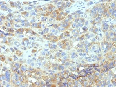
IHC, FFPE (verified)Validated Applications:
IHC, FFPEField of Research:
ImmunologyPositive Control:
Raji cells, MOLT 4, HeLa or SW480 cells. Human tonsil.Concentration:
0.1 mg/mLBuffer:
PBS, 0.1% BSA, 0.05% azideMolecular Weight:
85-115 kDaAdditionnal Information:
Immunohistology formalin-fixed 2-4 ug/mL|Staining of formalin-fixed tissues requires boiling tissue sections in 10 mM Tris with 1 mM EDTA, pH 9.0, for 10-20 min followed by cooling at RT for 20 minutes|Immunofluorescence 0.5-1 ug/mL|Flow Cytometry 0.5-1 ug/million cells/0.1 mL|Functional Studies order Ab without BSA & azide)|Optimal dilution for a specific application should be determined by userShipping Conditions:
Room temperatureStorage Conditions:
4°C; Stable at room temperature or 37°C (98°F) for 7 days.Shelf Life:
2 yearsCAS Number:
9007-83-4
DATASHEET Document
View DocumentMSDS Document
View Document