
Anti-CD50 (ICAM-3)(101-1D2), CF640R conjugate
CAT:
37-BNC400177-100
Size:
100 µL
Price:
Ask
- Availability: 24/48H Stock Items & 2 to 6 Weeks non Stock Items.
- Dry Ice Shipment: No


Anti-CD50 (ICAM-3)(101-1D2), CF640R conjugate
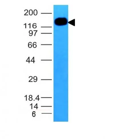
- Description: Recognizes an N-glycosylated glycoprotein of 120 kDa with intra-chain disulfide bonds, identified as CD50 or ICAM-3 (WS: IV & V). Its epitope localizes in the D2 extracellular domain and is resistant to neuraminidase and proteases. CD50 is the major ligand for LFA-1 (CD11a/CD18) and may have signalling role to increase adhesion. It is expressed on thymocytes and T lymphocytes and is resistant to treatment with phosphatidylinositol (PI) phospholipase C. This MAb inhibits primary mixed lymphocyte culture (MLC) but not secondary MLC, cytotoxicity or proliferation induced by mitogens. It blocks binding of NK1-L16 stimulated T cells to L cells expressing CD50. This MAb is excellent for staining of formalin/paraffin tissues.Primary antibodies are available purified, or with a selection of fluorescent CF® Dyes and other labels. CF® Dyes offer exceptional brightness and photostability. Note: Conjugates of blue fluorescent dyes like CF®405S and CF®405M are not recommended for detecting low abundance targets, because blue dyes have lower fluorescence and can give higher non-specific background than other dye colors.
- Synonyms: ICAMR; Intercellular adhesion molecule 3 (ICAM3)
- CAS Number: 9007-83-4
- UNSPSC: 41116161
- UNSPSC Description: Primary and secondary antibodies for multiple methodology immunostaining detection application
- Gene Name: ICAM3
- Gene ID: 3385
- NCBI Gene ID: 354563
- UniProt: P32942
- Cellular Locus: Plasma membrane
- Host: Mouse
- Species Reactivity: Human
- Immunogen: Stimulated human leukocytes
- Target Antigen: CD50
- Clonality: Monoclonal
- Isotype: IgG2a κ
- Clone: 101-1D2
- Conjugation: CF640R
- Source: Animal
- Applications: Flow, surface (published) | Functional studies (published) | ELISA (published)
- Validated Applications: FC, ELISA
- Field of Research: Immunology
- Positive Control: HL-60 or THP-1 cells. Lymph node and tonsil
- Concentration: 0.1 mg/mL
- Buffer: PBS, 0.1% BSA, 0.05% azide
- Molecular Weight: 110-160 kDa
- Additionnal Information: Higher concentration may be required for direct detection using primary antibody conjugates than for indirect detection with secondary antibody|Immunofluorescence: 0.5-1 ug/mL|Immunohistology formalin-fixed 0.5-1 ug/mL|Staining of formalin/paraffin tissues requires boiling tissue sections in 10 mM citrate buffer, pH 6.0, for 10-20 min followed by cooling at RT for 20 min|Flow Cytometry 0.5-1 ug/million cells/0.1 mL|Optimal dilution for a specific application should be determined by user
- References & Citations: Note: References for this clone sold by other suppliers may be listed for expected applications. Knapp, W. et. al. Leucocyte Typing IV, p541, 667-670, 1087, Oxford Univ. Press, 1989. Schlossman SF, et. al. Leucocyte Typing V, p1542-1547, 2011, Oxford Univ. Press, 1993. Eur J Immunol (1993) 23: 1508-1512. (IP, Flow) Eur J Immunol (1994) 24: 1377-1382. (functional studies) J of Immunol (1995) 154(6):3015-24. (ELISA)
- Shipping Conditions: Room temperature
- Storage Conditions: 4°C; Protect from light; Stable at room temperature or 37°C (98°F) for 7 days.
- Shelf Life: 2 years