
Anti-CD43(DF-T1), CF405S conjugate
CAT:
37-BNC040027-100
Size:
100 µL
Price:
Ask
- Availability: 24/48H Stock Items & 2 to 6 Weeks non Stock Items.
- Dry Ice Shipment: No


Anti-CD43(DF-T1), CF405S conjugate
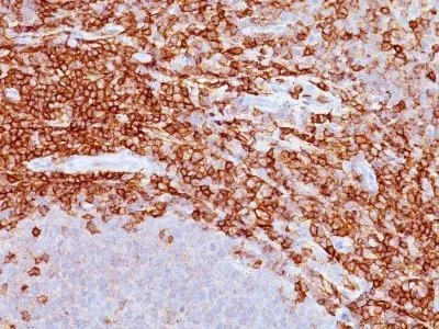
- Description: This antibody recognizes a cell surface glycoprotein of 95/115/135 kDa (depending upon the extent of glycosylation), identified as CD43 . Epitope of MAb Bra7G is clearly different from that of MAb DF-T1, called b as opposed to a for DF-T1. 70-90% of T-cell lymphomas and from 22-37% of B-cell lymphomas express CD43. No reactivity has been observed with reactive B-cells. So a B-lineage population that co-expresses CD43 is highly likely to be a malignant lymphoma, especially a low-grade lymphoma, rather than a reactive B-cell population. When CD43 antibody is used in combination with anti-CD20, effective immunophenotyping of the lymphomas in formalin-fixed tissues can be obtained. Co-staining of a lymphoid infiltrate with anti-CD20 and anti-CD43 argues against a reactive process and favors a diagnosis of lymphoma.Primary antibodies are available purified, or with a selection of fluorescent CF® Dyes and other labels. CF® Dyes offer exceptional brightness and photostability. Note: Conjugates of blue fluorescent dyes like CF®405S and CF®405M are not recommended for detecting low abundance targets, because blue dyes have lower fluorescence and can give higher non-specific background than other dye colors.
- Synonyms: Galactoglycoprotein, GALGP, GPL115, Leukocyte sialoglycoprotein, Leukosialin, LSN, Sialophorin, SPN
- CAS Number: 9007-83-4
- UNSPSC: 41116161
- UNSPSC Description: Primary and secondary antibodies for multiple methodology immunostaining detection application
- Gene Name: SPN
- Gene ID: 6693
- NCBI Gene ID: 632188
- UniProt: P16150
- Cellular Locus: Plasma membrane
- Host: Mouse
- Species Reactivity: Human
- Immunogen: Myeloblastic KG1 cells
- Target Antigen: CD43
- Clonality: Monoclonal
- Isotype: IgG1 κ
- Clone: DF-T1
- Conjugation: CF405S
- Disease: Tumor
- Source: Animal
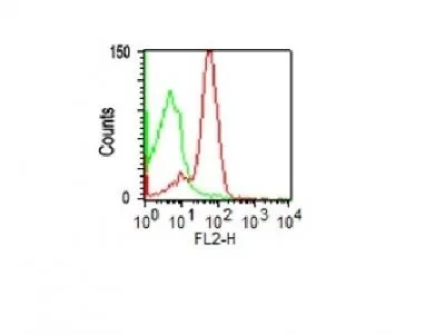
- Applications: Flow (verified) | IF (verified) | IHC, FFPE (verified) | WB (verified)
- Validated Applications: FC, IF, IHC, FFPE, WB
- Field of Research: Immunology
- Positive Control: Paracortex in a tonsil or a reactive lymph node
- Concentration: 0.1 mg/mL
- Buffer: PBS, 0.1% BSA, 0.05% azide
- Molecular Weight: 95, 115, or 135 kDa
- Additionnal Information: Higher concentration may be required for direct detection using primary antibody conjugates than for indirect detection with secondary antibody|Immunofluorescence: 1-2 ug/mL|Immunohistology formalin-fixed 0.5-1 ug/mL|Staining of formalin-fixed tissues requires boiling tissue sections in 10 mM citrate buffer, pH 6.0, for 10-20 min followed by cooling at RT for 20 minutes|Flow Cytometry 0.5-1 ug/million cells/0.1 mL|Optimal dilution for a specific application should be determined by user
- Shipping Conditions: Room temperature
- Storage Conditions: 4°C; Protect from light; Stable at room temperature or 37°C (98°F) for 7 days.
- Shelf Life: 2 years