Anti-bcl-x(BX006)
CAT:
37-BNUM0006-50
Size:
50 µL
Price:
Ask
- Availability: 24/48H Stock Items & 2 to 6 Weeks non Stock Items.
- Dry Ice Shipment: No


Anti-bcl-x(BX006)
Description:
Bcl-x belongs to the Bcl-2 family of proteins and plays a crucial role in apoptosis. Bcl-X has two isoforms: Bcl-XL (long), a 26 kDa protein which suppresses cell death, and Bcl-XS (short), a 178 amino acid protein lacking a 63 amino acid domain which functions as a dominant inhibitor of Bcl-2 and promotes apoptosis. This MAb reacts with both Bcl-XS and Bcl-XL proteins, and shows no cross-reaction with Bcl-2 or Bax proteins.Primary antibodies are available purified, or with a selection of fluorescent CF® Dyes and other labels. CF® Dyes offer exceptional brightness and photostability. Note: Conjugates of blue fluorescent dyes like CF®405S and CF®405M are not recommended for detecting low abundance targets, because blue dyes have lower fluorescence and can give higher non-specific background than other dye colors._x000D_ _x000D_Synonyms:
Bcl-x; Bcl-2-like protein 1; BCL2L1; Apoptosis regulator Bcl-XUNSPSC:
41116161UNSPSC Description:
Primary and secondary antibodies for multiple methodology immunostaining detection applicationGene Name:
BCL2L1Gene ID:
598NCBI Gene ID:
516966UniProt:
Q07817Cellular Locus:
Cytoplasmic|Endoplasmic reticulum|MitochondriaHost:
MouseSpecies Reactivity:
Human, Mouse, Pig, RatImmunogen:
A synthetic peptide, aa 3-14 (Cys-QSNRELVVDFLS) of human Bcl-X protein.Target Antigen:
Bcl-xClonality:
MonoclonalIsotype:
IgG2aClone:
BX006Conjugation:
Purified, BSA-freeSource:
AnimalApplications:
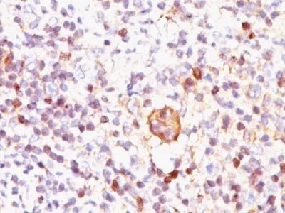
IHC, FFPE (verified) | WB (verified)Validated Applications:
IHC, FFPE, WBField of Research:
Apoptosis, CancerPositive Control:
Jurkat, K562, HL-60, or HeLa Cells. Reed-Sternberg cells in Hodgkin's lymphoma.Concentration:
1 mg/mLBuffer:
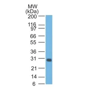
PBS, no BSA, no azideMolecular Weight:
27 kDaAdditionnal Information:
Higher concentration may be required for direct detection using primary antibody conjugates than for indirect detection with secondary antibody|Immunofluorescence: 1-2 ug/mL|Immunohistology formalin-fixed 0.5-1 ug/mL|Staining of formalin-fixed tissues requires boiling tissue sections in 10 mM Tris with 1 mM EDTA, pH 9.0, for 10-20 min followed by cooling at RT for 20 minutes|Flow Cytometry 0.5-1 ug/million cells/0.1 mL|Western blotting 0.5-1 ug/mL|Optimal dilution for a specific application should be determined by userShipping Conditions:
Room temperatureStorage Conditions:
-35°C to -5°C ; Stable at room temperature or 37°C (98°F) for 7 days.Shelf Life:
2 yearsCAS Number:
9007-83-4
DATASHEET Document
View DocumentMSDS Document
View Document