Anti-BCL-10(BL10/411)
CAT:
37-BNUM0411-50
Size:
50 µL
Price:
Ask
- Availability: 24/48H Stock Items & 2 to 6 Weeks non Stock Items.
- Dry Ice Shipment: No


Anti-BCL-10(BL10/411)
Description:
B-cell lymphoma/leukemia 10 (BCL10) is a signaling protein that promotes apoptosis, pro-caspase-9 maturation, and activation of NF-kappa-B via NIK and IKK. BCL10 contains an N-terminal caspase recruitment domain (CARD), which is found in a number of apoptotic regulatory molecules. BCL10 is involved in the adaptive immune response, and may be an adapter protein between upstream TNFR1-TRADD-RIP complex and the downstream NIK-IKK-IKAP complex. A BCL10 translocation is recurrent in low-grade mucosa-associated lymphoid tissue (MALT lymphoma)Primary antibodies are available purified, or with a selection of fluorescent CF® Dyes and other labels. CF® Dyes offer exceptional brightness and photostability. Note: Conjugates of blue fluorescent dyes like CF®405S and CF®405M are not recommended for detecting low abundance targets, because blue dyes have lower fluorescence and can give higher non-specific background than other dye colors.Synonyms:
BCL10; BCL-10; B-cell CLL/lymphoma 10; B-cell leukemia/lymphoma 10UNSPSC:
41116161UNSPSC Description:
Primary and secondary antibodies for multiple methodology immunostaining detection applicationGene Name:
BCL10Gene ID:
8915NCBI Gene ID:
193516UniProt:
O95999Cellular Locus:
Nucleus & cytoplasmHost:
MouseSpecies Reactivity:
HumanImmunogen:
Human BCL10 recombinant protein (epitope aa122-168)Target Antigen:
BCL10Clonality:
MonoclonalIsotype:
IgG1 κClone:
BL10/411Conjugation:
Purified, BSA-freeSource:
AnimalApplications:
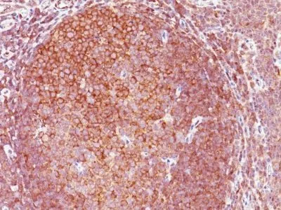
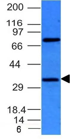
Flow, intracellular (verified) | IHC, FFPE (verified) | WB (verified)Validated Applications:
FC, IHC, FFPE, WBField of Research:
Apoptosis, Cancer, Immunology, Signal transductionPositive Control:
HepG2 cells or Lymphoma.Concentration:
1 mg/mLBuffer:
PBS, no BSA, no azideMolecular Weight:
33 kDaAdditionnal Information:
Higher concentration may be required for direct detection using primary antibody conjugates than for indirect detection with secondary antibody|Immunofluorescence: 1-2 ug/mL|Immunohistology formalin-fixed 0.5-1 ug/mL|Staining of formalin-fixed tissues requires boiling tissue sections in 10 mM citrate buffer, pH 6.0, for 10-20 min followed by cooling at RT for 20 minutes|Flow Cytometry 0.5-1 ug/million cells/0.1 mL|Western blotting 0.5-1 ug/mL|Optimal dilution for a specific application should be determined by userShipping Conditions:
Room temperatureStorage Conditions:
-35°C to -5°C ; Stable at room temperature or 37°C (98°F) for 7 days.Shelf Life:
2 yearsCAS Number:
9007-83-4
DATASHEET Document
View DocumentMSDS Document
View Document