
Anti-Bax(BAX/962)
CAT:
37-BNUB0962-100
Size:
100 µL
Price:
Ask
- Availability: 24/48H Stock Items & 2 to 6 Weeks non Stock Items.
- Dry Ice Shipment: No


Anti-Bax(BAX/962)
- Description: This antibody recognizes a protein of 21 kDa, identified as the Bax protein. This MAb is highly specific to Bax and shows no cross-reaction with Bcl-2 or Bcl-X protein. Bcl-2 blocks cell death following a variety of stimuli. Bax has extensive amino acid homology with Bcl-2 and it homodimerizes and forms heterodimers with Bcl-2. Overexpression of Bax accelerates apoptotic death induced by cytokine deprivation in an IL-3 dependent cell line, and Bax also counters the death repressor activity of Bcl-2._x000D__x000D_Primary antibodies are available purified, or with a selection of fluorescent CF® Dyes and other labels. CF® Dyes offer exceptional brightness and photostability. Note: Conjugates of blue fluorescent dyes like CF®405S and CF®405M are not recommended for detecting low abundance targets, because blue dyes have lower fluorescence and can give higher non-specific background than other dye colors._x000D_ _x000D_
- Synonyms: BAX; Apoptosis regulator BAX; BCL2 associated X protein; Bcl-2-like protein 4
- CAS Number: 9007-83-4
- UNSPSC: 41116161
- UNSPSC Description: Primary and secondary antibodies for multiple methodology immunostaining detection application
- Gene Name: BAX
- Gene ID: 581
- NCBI Gene ID: 624291
- UniProt: Q07812
- Cellular Locus: Mitochondria
- Host: Mouse
- Species Reactivity: Human, Monkey
- Immunogen: Recombinant full-length human BAX protein.
- Target Antigen: Bax
- Clonality: Monoclonal
- Isotype: IgG1
- Clone: BAX/962
- Conjugation: Purified, with BSA
- Source: Animal
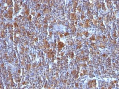
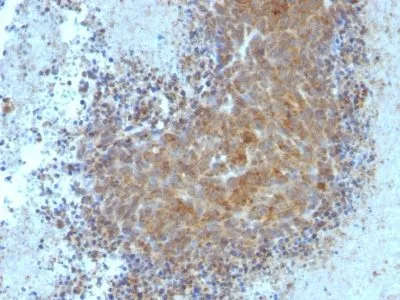
- Applications: Flow, intracellular (verified) | IHC, FFPE (verified)
- Validated Applications: FC, IHC, FFPE
- Field of Research: Apoptosis, Cancer
- Positive Control: Jurkat, K562, HL-60, or HeLa Cells. Reed-Sternberg cells in Hodgkin's lymphoma.
- Concentration: 0.2 mg/mL
- Buffer: PBS, 0.05% BSA, 0.05% azide
- Molecular Weight: 21 kDa
- Additionnal Information: Higher concentration may be required for direct detection using primary antibody conjugates than for indirect detection with secondary antibody|Immunofluorescence: 1-2 ug/mL|Does not react with mouse or rat, others not known|Immunohistology formalin-fixed 0.5-1 ug/mL|Staining of formalin-fixed tissues requires boiling tissue sections in 10 mM Tris with 1 mM EDTA, pH 9.0, for 10-20 min followed by cooling at RT for 20 minutes|Flow Cytometry 0.5-1 ug/million cells/0.1 mL|Western blotting 0.5-1 ug/mL|Optimal dilution for a specific application should be determined by user
- Shipping Conditions: Room temperature
- Storage Conditions: 4°C; Stable at room temperature or 37°C (98°F) for 7 days.
- Shelf Life: 2 years