
Anti-AFP (Alpha Fetoprotein)(C3)
CAT:
37-BNUB0354-500
Size:
500 µL
Price:
Ask
- Availability: 24/48H Stock Items & 2 to 6 Weeks non Stock Items.
- Dry Ice Shipment: No


Anti-AFP (Alpha Fetoprotein)(C3)
- Description: Alpha-fetoprotein (AFP) is an oncofetal glycoprotein with a single chain of 70 kDa. This MAb is highly specific to AFP and shows no cross-reaction with other oncofetal antigens or serum albumin. AFP is normally synthesized in the liver, intestinal tract, and yolk sac of the fetus. Antibody to AFP has been shown to be useful in detecting hepatocellular carcinomas (HCC) and germ cell neoplasms, especially yolk sac tumors.Primary antibodies are available purified, or with a selection of fluorescent CF® Dyes and other labels. CF® Dyes offer exceptional brightness and photostability. Note: Conjugates of blue fluorescent dyes like CF®405S and CF®405M are not recommended for detecting low abundance targets, because blue dyes have lower fluorescence and can give higher non-specific background than other dye colors.
- Synonyms: Alpha-fetoglobulin; FETA; HPAFP
- CAS Number: 9007-83-4
- UNSPSC: 41116161
- UNSPSC Description: Primary and secondary antibodies for multiple methodology immunostaining detection application
- Gene Name: AFP
- Gene ID: 174
- NCBI Gene ID: 518808
- UniProt: P02771
- Cellular Locus: Cytoplasmic|Golgi apparatus
- Host: Mouse
- Species Reactivity: Dog, Human, Monkey, Pig
- Immunogen: Alpha fetoprotein (AFP) purified from serum of a hepatoma patient
- Target Antigen: AFP | Alpha-fetoprotein
- Clonality: Monoclonal
- Isotype: IgG2a κ
- Clone: C3
- Conjugation: Purified, with BSA
- Disease: Tumor
- Source: Animal
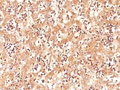
- Applications: IHC, FFPE (verified)
- Validated Applications: IHC, FFPE
- Field of Research: Cancer, Developmental biology
- Positive Control: Hep-G2 cells. Fetal liver or hepatocellular carcinoma
- Concentration: 0.2 mg/mL
- Buffer: PBS, 0.05% BSA, 0.05% azide
- Molecular Weight: 70 kDa
- Additionnal Information: Higher concentration may be required for direct detection using primary antibody conjugates than for indirect detection with secondary antibody|Immunofluorescence: 0.5-1 ug/mL|Immunohistology formalin-fixed 1-2 ug/mL|Does not react with cow, dog, mouse, or rat|Staining of formalin-fixed tissues requires boiling tissue sections in 10 mM citrate buffer, pH 6.0, for 10-20 min followed by cooling at RT for 20 minutes|Flow Cytometry 0.5-1 ug/million cells/0.1 mL|Optimal dilution for a specific application should be determined by user
- Shipping Conditions: Room temperature
- Storage Conditions: 4°C; Stable at room temperature or 37°C (98°F) for 7 days.
- Shelf Life: 2 years