Products for Research Use Only

Cell Cycle Assay Kit (Red Fluorescence)
CAT: 0763-E-CK-A351-01Size: 20 AssaysDry Ice: NoHazardous: No
Product image 1
1 / 1
CAT#:0763-E-CK-A351-01Size:20 Assays
Selected
24/48H Stock Items & 2 to 6 Weeks non Stock Items.
Quick Request Actions
Detection Method
Fluorometric method; Flow cytometry
Assay Principle
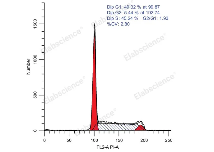
Cell cycle refers to the whole process from the end of one mitosis to the end of the next. During this process, the genetic material is replicated and doubled, and evenly distributed to two daughter cells at the end of division. Cell cycle can also be divided into phases like interphase and Metaphase. Intercellular phase can also be divided into dormancy (zero gap, G0), prophase of DNA synthesis (first gap, G1), anaphase of DNA synthesis (synthesis, S) and anaphase of DNA synthesis (second gap, G2) . DNA detection can be used to reflect the status of each phase of the cell cycle which is also named cell proliferation. DNA can bind to fluorescent dyes (such as propidium Iodide-PI), the fluorescent dyes binding to DNA at different stages are different, and the fluorescence intensity detected by flow cytometry can also be used to detect different phases in cell cycle. When apoptotic cells occur, apoptotic bodies are produced due to the concentration and nucleolysis of cytoplasm and chromatin, which will change the light scattering properties of cells. In the early stage of apoptosis, the forward scattering (FSC) of cells decreased significantly, while the side scatter (SSC) increased or remained unchanged. In the late stage of apoptosis, both FSC and SSC signals decreased. Therefore, apoptotic cells can be observed by measuring the changes of light scattering by flow cytometry. After staining with PI, assuming that the fluorescence intensity of G0/G1 phase cells is 1, the theoretical value of fluorescence intensity of G2/M phase cells containing double genomic DNA is 2, and the fluorescence intensity of S phase cells undergoing DNA replication is between 1 and 2. Apoptotic cells lost part of genomic DNA fragmentation due to nucleus concentration and DNA fragmentation. Therefore, apoptotic cells showed obvious weak staining after PI staining and the fluorescence intensity was less than 1. The sub-G1 peak appeared on the flow cytometry fluorescence map which is apoptotic cell. The kit can be used to detect the DNA content (cell cycle) of cultured cells (suspension or adherence cells) .
Sample Type
Cell samples
Shipping Conditions
Ice bag
Storage Conditions
This product can be stored at 2~8°C/-20°C for 12 months with shading light. Please refer to the manual for the specific storage condition of the components.
Citation 01
https://www.sciencedirect.com/science/article/pii/S2452199X25001720
Citation 02
https://onlinelibrary.wiley.com/doi/abs/10.1002/adfm.202408511
Citation 03
https://www.nature.com/articles/s41467-024-46141-x
Citation 04
https://onlinelibrary.wiley.com/doi/abs/10.1002/adhm.202401398
Citation 05
https://advanced.onlinelibrary.wiley.com/doi/abs/10.1002/adhm.202402884
Citation 06
https://www.sciencedirect.com/science/article/pii/S0168365924009283
Citation 07
https://www.ncbi.nlm.nih.gov/pmc/articles/PMC8741845/
Citation 08
https://www.sciencedirect.com/science/article/pii/S0043135420312884
Citation 09
https://aacrjournals.org/cancerimmunolres/article/doi/10.1158/2326-6066.CIR-24-0039/749533
Citation 10
https://www.nature.com/articles/s41419-025-07713-x
Citation 11
https://spj.science.org/doi/abs/10.34133/bmr.0194
Citation 12
https://www.sciencedirect.com/science/article/pii/S2162253120303358
Detection Instrument
Flow Cytometer