Intracellular Total ROS Activity Assay
CAT:
436-9144
Size:
100 Tests
Price:
Ask
- Availability: 24/48H Stock Items & 2 to 6 Weeks non Stock Items.
- Dry Ice Shipment: No


Intracellular Total ROS Activity Assay
Description:
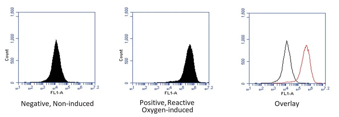
The Intracellular Total ROS Activity Assay enables the screening of oxidative stress inhibitor and activator reagents for potency. The assay assesses the overall level of intracellular ROS activity. Analyze using a flow cytometer.Target:
ROSLabel:
ICTCell Type:
JurkatType:
Cell ViabiityDetection Method:
Flow cytometryWavelength:
490 nm / 520 nmAssay Protocol:
1. Prepare samples and controls., 2. Dilute 10X Cellular Assay Buffer 1:10 with diH2O., 3. Reconstitute FAM-FLICA with 50 µL DMSO., 4. Dilute FAM-FLICA 1:5 by adding 200 µL PBS., 5. Add diluted FAM-FLICA to each sample at 1:30-1:60 (e.g., spike at 1:30 by adding 10 µL to 290 µL sample)., 6. Incubate approximately 1 hour., 7. Remove media and wash cells 3 times: add 1X Cellular Wash Buffer and spin cells., 8. If desired, label with additional stains, such as Hoechst 33342, Propidium Iodide, 7-AAD, or an antibody., 9. If desired, fix cells., 10. Analyze with a fluorescence microscope, fluorescence plate reader, or flow cytometer. FAM-FLICA excited at 492 nm and emits at 520 nm.Sample Type:
Cell cultureComponents:
Total ROS Green, lyophilized, 1 vial, #6688, DMSO, 200 µL, 1 vial, #6689, Assay Buffer, 10X, 15 mL, #686, Kit ManualShipping Conditions:
Ships overnight (domestic), International Priority ShippingStorage Temperature:
Total ROS Green and DMSO at ≤-20°C, Assay Buffer at ≤2-8°CNotes:
20% discountCellular Imaging & Detection:
Oxidative StressTarget Description:
Reactive oxygen species (ROS) are natural byproducts of the normal metabolism of oxygen and play important roles in cell signaling. How- ever, during oxidative stress-related states, ROS levels can increase dramatically. The accumulation of ROS results in significant damage to cellular structures. The role of oxidative stress in cardiovascular disease, diabetes, osteoporosis, stroke, inflammatory diseases, a number of neuro-degenerative diseases and cancer, has been Well established. ICT’s Intracellular Total ROS Activity Assay provides a good screening option for assessing the potency of oxidative stress inhibitor and activator reagents, and will help to determine how oxidative stress modulates varied intracellular pathways. This kit assesses the overall level of intracellular ROS activity, but does not identify the specific reactive oxygen molecule(s) generated by the oxidative stress event. The kit provides all the essential reagents and an easy to follow protocol to assess changes in intracellular ROS levels by flow cytometry. The key reagent in the assay is a proprietary dye called Total ROS Green. This dye quickly penetrates membrane structures and accumulates within the cell. In the presence of ROS, the non-fluorescent Total ROS Green dye molecule is oxidized by all various iterations of ROS molecular forms. In the oxidized state, the Total ROS Green dye molecule acquires fluorescence properties that enable its detection by flow cytometry (Ex/Em: 490 nm/520 nm) as an indicator of the relative level of intracellular ROS activity. This assay is not designed to determine which particular cell pathology triggered the increase in intracellular ROS concentration in the treatment cell populations, or which individual species of reactive oxygen are involved in the cell response. Other testing parameters must be analyzed to establish the specific causation process responsible for the increased intracellular ROS activity levels. ICT’s Intracellular Total ROS Activity Assay requires minimal procedural steps and hands-on time to complete. Suspension cells or EDTA disassociated adherent cells are stained for 1 hour to pre-load the cells with Total ROS Green quantitation dye. Cells are then treated experimentally. Since the non-internalized dye probe is non-fluorescent, subsequent wash steps are not required, thus simplifying the assay procedure. Following treatment, cells are ready for analysis by flow cytometry. Each kit will enable the assessment of up to 100 samples (0.5 mL each). This assay can be adapted for use with a fluorescence microscope or plate reader equipped with FITC/FAM dye-compatible excitation/emission optics. Use and optimization of these alternative fluorescence analysis methods will require further modification of this flow cytometer based protocol.