Anti-Tyrosinase-Related Protein-1 (TYRP-1) (Melanoma Marker) (TYRP1/2340R)
CAT:
37-BNUM2340-50
Size:
50 µL
Price:
Ask
- Availability: 24/48H Stock Items & 2 to 6 Weeks non Stock Items.
- Dry Ice Shipment: No


Anti-Tyrosinase-Related Protein-1 (TYRP-1) (Melanoma Marker) (TYRP1/2340R)
Description:
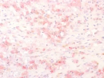
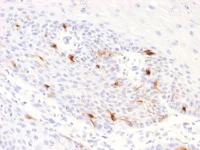
This antibody reacts with a 75 kDa melanocyte-specific gene product, identified as Tyrosinase-Related Protein-1 (TRP-1). It is involved in melanin synthesis. TRP1 is present on the melanosomal membranes of melanoma, normal melanocytes and nevi. Recent evidence suggests that TRP-1 is involved in maintaining stability of tyrosinase protein and modulating its catalytic activity. TRP-1 is also involved in maintenance of melanosome ultrastructure and affects melanocyte proliferation and cell death.Primary antibodies are available purified, or with a selection of fluorescent CF® Dyes and other labels. CF® Dyes offer exceptional brightness and photostability. Note: Conjugates of blue fluorescent dyes like CF®405S and CF®405M are not recommended for detecting low abundance targets, because blue dyes have lower fluorescence and can give higher non-specific background than other dye colors.Synonyms:
5; 6 dihydroxyindole 2 carboxylic acid oxidase; 6-dihydroxyindole-2-carboxylic acid oxidase; Associated with iris pigmentation; CAS2; Catalase B (CATB); DHICA oxidase; Glycoprotein75 (GP75); Melanoma antigen gp75; Tyrosinase-related protein 1 (TYRP1); TYRRPUNSPSC:
41116161UNSPSC Description:
Primary and secondary antibodies for multiple methodology immunostaining detection applicationGene Name:
TYRP1Gene ID:
7306NCBI Gene ID:
270279UniProt:
P17643Cellular Locus:
VesicularHost:
RabbitSpecies Reactivity:
HumanImmunogen:
Recombinant fragment of human TYRP1 protein (around aa 257-377) (exact sequence is proprietary)Target Antigen:
Tyrosinase Related Protein-1Clonality:
Recombinant MonoclonalIsotype:
IgGClone:
TYRP1/2340RConjugation:
Purified, BSA-freeDisease:
TumorSource:
AnimalApplications:
IHC, FFPE (verified)Validated Applications:
IHC, FFPEField of Research:
CancerPositive Control:
SK-MEL-23, SK-MEL-19, SK-MEL-30 cells. Human Skin or Melanoma.Concentration:
1 mg/mLBuffer:
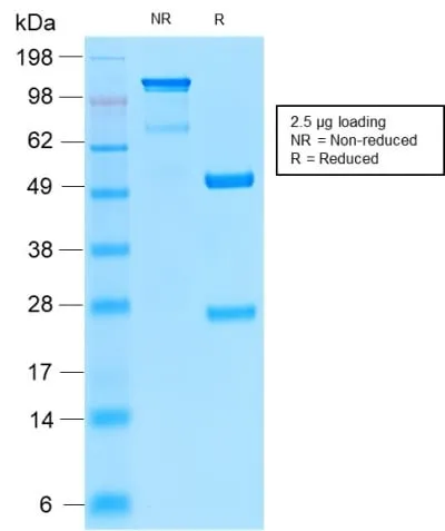
PBS, no BSA, no azideMolecular Weight:
75 kDaAdditionnal Information:
Higher concentration may be required for direct detection using primary antibody conjugates than for indirect detection with secondary antibody|ELISA: 2-4 ug/m for coating order Ab without BSA|Immunohistology (formalin): 1-2 ug/mL for 30 minutes at RT|Staining of formalin-fixed tissues requires boiling tissue sections in 10 mM citrate buffer, pH 6.0, for 10-20 minutes followed by cooling at RT for 20 minutes|Optimal dilution for a specific application should be determined by userShipping Conditions:
Room temperatureStorage Conditions:
-35°C to -5°C ; Stable at room temperature or 37°C (98°F) for 7 days.Shelf Life:
2 yearsCAS Number:
9007-83-4
DATASHEET Document
View DocumentMSDS Document
View Document